Flexible Detector Bundle

Imagent provides a balance between temporal and spatial resolution for the cognitive study of superficially located areas of human brain by addressing two main applications techniques:
Brain imaging techniques can be broadly classified in two groups. One group includes the techniques that have a good spatial resolution (up to 1 - 2 mm) but a poor temporal resolution, such as functional Magnetic Resonance Imaging (fMRI) and Positron Emission Tomography (PET). The second group includes techniques featuring an excellent temporal resolution (of the order of milliseconds) but providing a limited spatial information. This group includes the Event Related Brain Potential (ERP) and the Magneto-encephalography (MEG). Imagent captures both the slow signals (hemodynamic changes) and the fast signals (EROS).
Notice: Investigational device. Limited by Federal (or United States) law to investigational use. The ISS Imagent is presently used for research only.

Measures Both Amplitude & Changes in Average Transit Time

Fast Measurements Up to 50 Hz

Applicable to a Wide Range of Research
Cognitive Neuroscience
Physiological monitoring
Virtual Reality
Brain Computer Interface
Cognitive Neuroscience
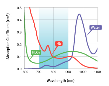
Imagent's working principle is based on the use of near infrared light for probing the cortical surface. The main tissue absorbers in the wavelength region spanning from 700 nm to 900 nm are oxy-hemoglobin (HbO2) and deoxy-hemoglobin (Hb); on a smaller scale, water, fat and cytochrome oxidase contribute to the partial absorption of the light. The penetration depth of light in tissues is quite significant in this wavelength range. For typical head tissue (skin/scalp, skull and cortical layer), with an absorption coefficient of μa = 0.1 cm-1 and a scattering coefficient μs' = 8 cm-1, the maximum optical penetration can be estimated to be about 1.5 cm when a detector is placed at 4 cm from the source. The penetration depth can be increased by increasing the distance between the source and the detector, although, eventually, the signal-to-noise ratio of the measurement deteriorates.
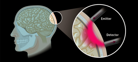
Imagent utilizes laser diodes emitting at 690 nm and 830 nm. The light is delivered by fiber optics positioned on the head. Upon entering the tissue, the near infrared light, albeit weakly absorbed, is highly scattered by the tissue in homogeneities. A fraction of the light leaves the tissue and it is collected by the collecting fiber that carries it back to the light detectors housed in the unit for data processing. The fibers are kept in place by a headgear, which is available for adults and children; for the study of specific areas, pad sensors are available. Up to 128 fibers and up to 60 detectors bundles (for a total of 3,840 optical channels) can be positioned on the head of an adult. Different patterns (montages) of the excitation and collection fibers can be used.
Imagent utilizes the frequency domain technology; whereas, the light sources are modulated at high frequency (of the order of 100 MHz) and three parameters of the detected signal are measured: the intensity, the modulation depth and the time it takes to traverse the tissue (phase delay). Any two combinations of the three measured quantities can be utilized to provide changes in the physiological parameters, the choice being dictated by the specific parameter to be measured, by the need to reduce physiological noise and by the time scale of the event to be measured.