NEWS
PLIM Acquisition Implemented on the Nikon A1 Multi Photon Microscope System
Champaign, Illinois - November 30, 2021 - Phosphorescence has a number of promising applications in biomedical research. Phosphorescent probes can be engineered to respond to environmental parameters, such as tissue O2 concentration. These probes typically have decay lifetimes in the range of micro- to milliseconds, as compared to fluorescence probes that have lifetimes on the order of nanoseconds. Therefore, measurements of phosphoresce lifetimes require excitation light sources operating at low repetition rates.
Previously, ISS introduced a phosphorescence lifetime imaging (PLIM) module compatible with excitation sources having repetition rates as low as 1 Hz. Recently, ISS developed a new module that enables PLIM using Ti-Sapphire femtosecond oscillators operating at repetition rates of 80-100 MHz, which are typically used in two-photon (2P) microscopy. This new module has been combined with Nikon A1 2-photon microscope and shown capable of lifetime measurements in the range of 100 ns to 100 ms.
Oxygen quenches phosphorescence of probe Oxyphor 2P [1] (kindly provided by the center for Oxygen Imaging by Phosphorescence Quenching, NIH grant U24-EB028941, PI: Dr. Sergei Vinogradov, University of Pennsylvania). When this probe is injected into tissue, oxygen concentration can be imaged with micron-scale resolution using the method of two-photon-excited phosphorescence [2-4] at depths reaching 600µm below the tissue surface. As a demonstration of principle, the probe was injected systemically in a mouse under anesthesia, and the oxygen concentration in the inhaled gas mixture was varied between 15% and 100%. Two-photon PLIM in the brain cortex was performed through a cranial window using a laser emitting at 960 nm. The phosphorescence was detected using a GaAs PMT and a FastFLIM card. The decay times of Oxyphor 2P vary in the range of ~9-38 µs, quantitatively reflecting changes in the brain tissue oxygenation. All institutional IACUC guidelines were followed during the procedure. [Funded by the grant NS083858 from the NIH (PI: Dr. Sergei Kirov, Medical College of Georgia at Augusta University).]
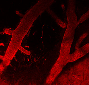
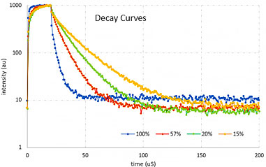
Figures courtesy of Dr. J. Sword, Medical College of Georgia at Augusta University Medical Center, Augusta, GA
ISS Nikon A1 FLIM & PLIM Upgrades Kits provide solutions for:
- FFS (FCS, PCH, RICS, and N&B)
- Scanning FCS
- FLIM/FRET
- PLIM
- Orbital Particle Tracking
References
- Esipova, T. V.; Barrett, M. J. P.; Erlebach, E.; Masunov, A. E.; Weber, B.; Vinogradov, S. A.;
Oxyphor 2P: A High-Performance Probe for Deep-Tissue Longitudinal Oxygen Imaging.
Cell Metabolism 2019, 29 (3), 736-744. - Finikova, O. S.; Lebedev, A. Y.; Aprelev, A.; Troxler, T.; Gao, F.; Garnacho, C.; Muro, S.; Hochstrasser, R. M.; Vinogradov, S. A.;
Oxygen Microscopy by Two-Photon-Excited Phosphorescence.
ChemPhysChem 2008, 9 (12), 1673-1679. - Sakadzic, S.; Roussakis, E.; Yaseen, M. A.; Mandeville, E. T.; Srinivasan, V. J.; Arai, K.; Ruvinskaya, S.; Devor, A.; Lo, E. H.; Vinogradov, S. A., Boas D. A.;
Two-Photon High-Resolution Measurement of Partial Pressure of Oxygen in Cerebral Vasculature and Tissue.
Nature Methods 2010, 7 (9), 755-U125. - Lecoq, J.; Parpaleix, A.; Roussakis, E.; Ducros, M.; Houssen, Y. G.; Vinogradov, S. A.; Charpak, S.;
Flow Simultaneous Two-Photon Imaging of Oxygen and Blood in Deep Cerebral Vessels.
Nature Medicine 2011, 17 (7), 893-U262.