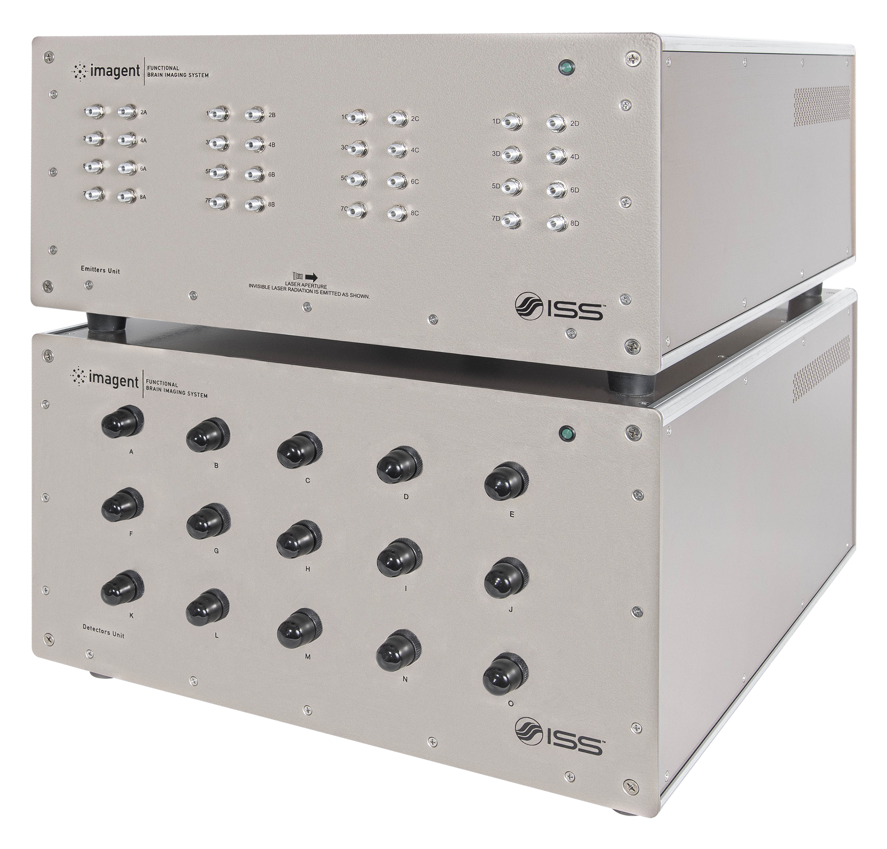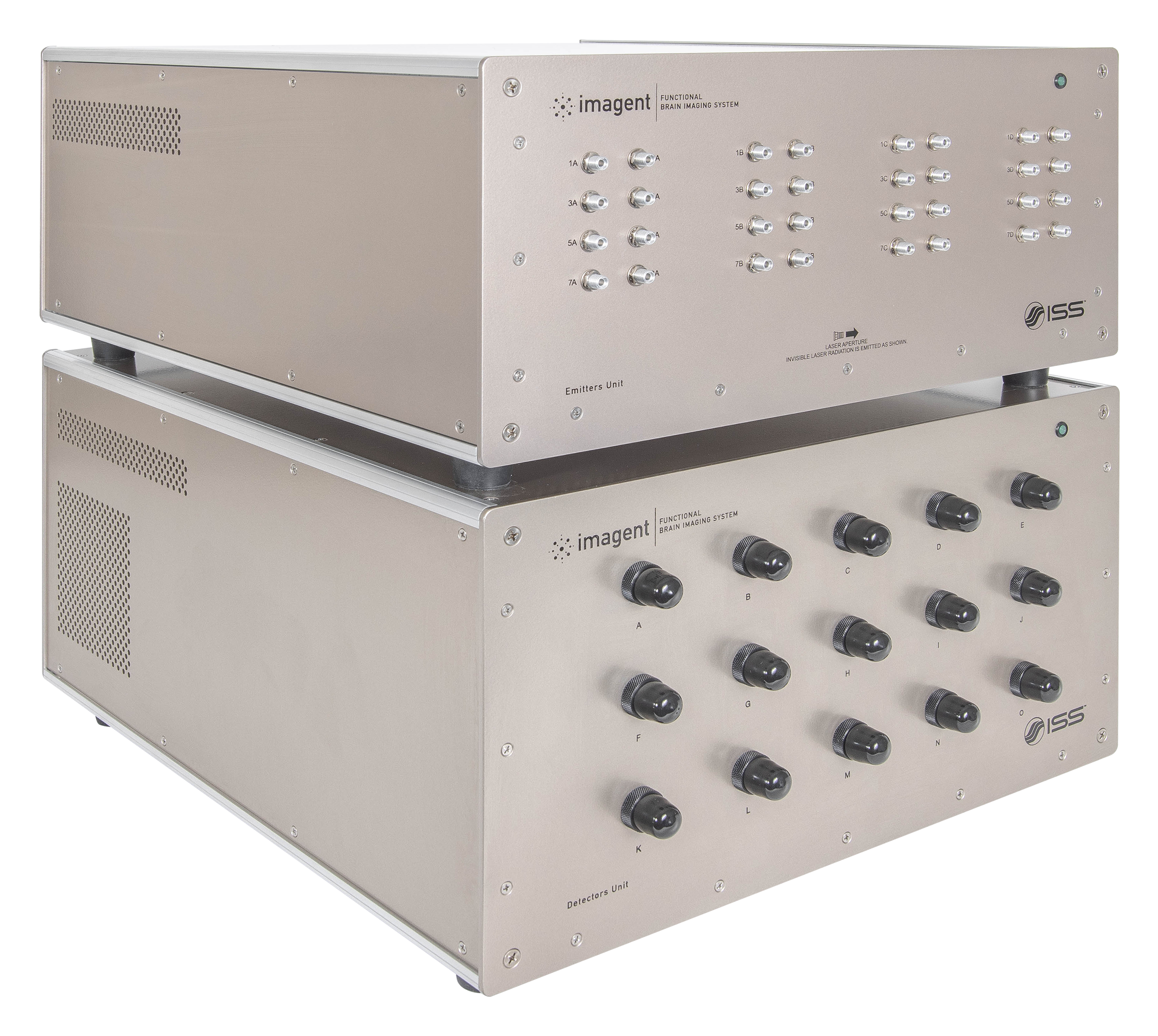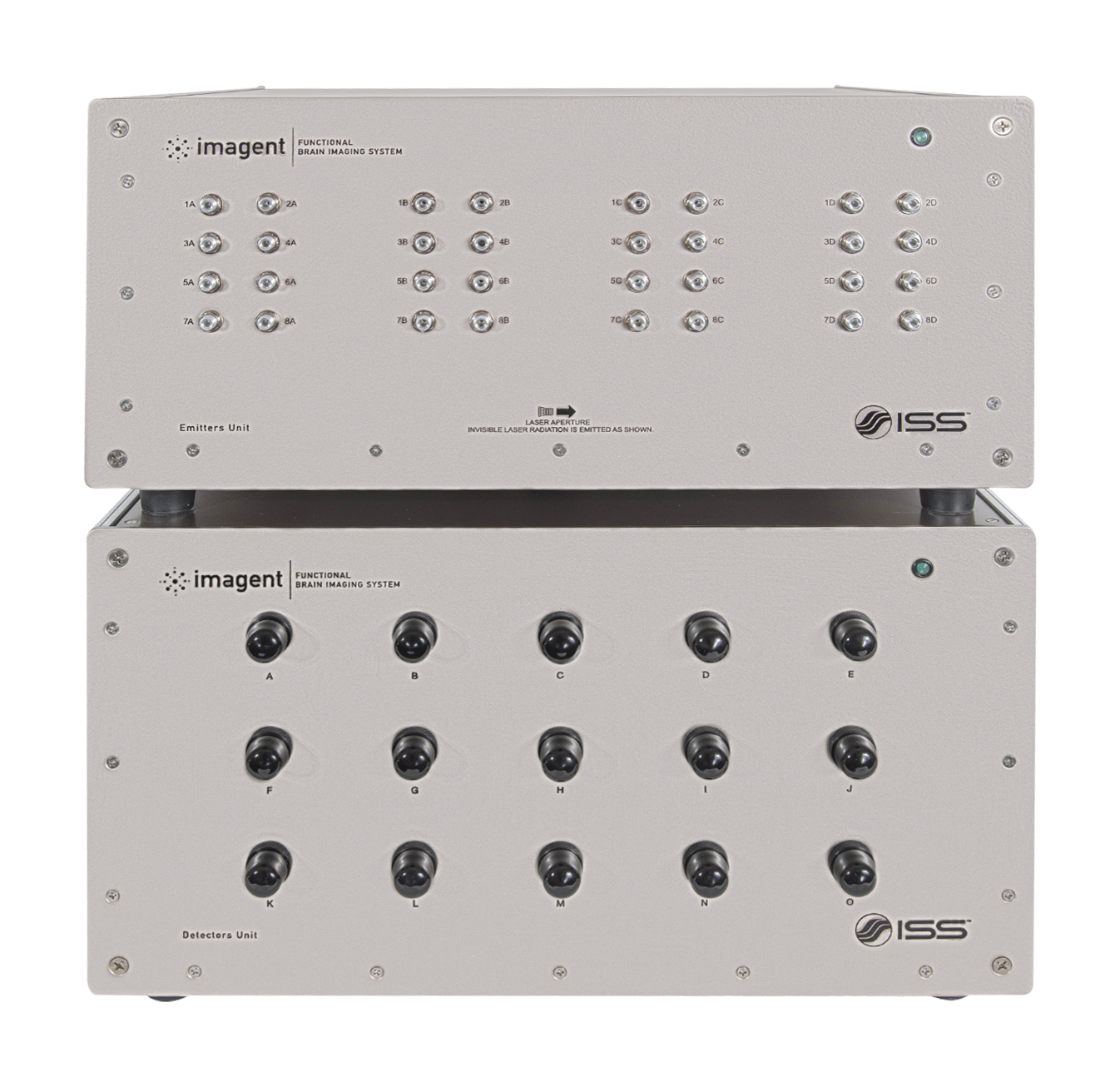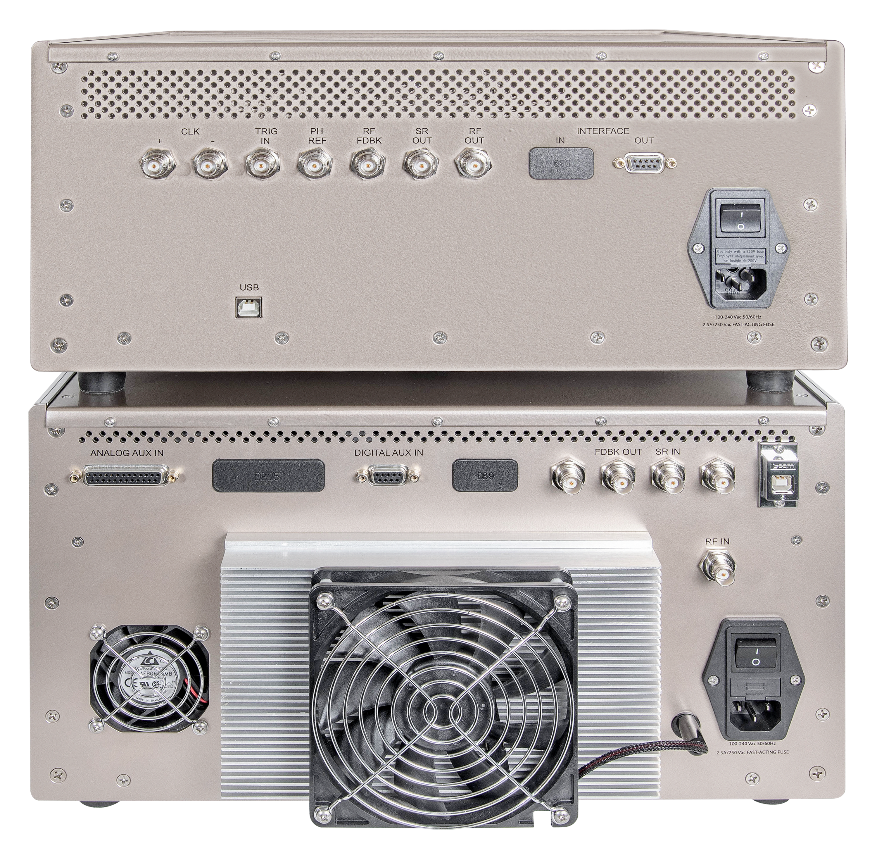
Overview of Imagent
Imagent为人脑表面区域相关的认知学研究提供了时间与空间分辨率之间的平衡,主要通过以下两个技术来实现:
- 功能性近红外光谱技术 (fNIRS):这项技术探测了外界刺激下光学信号的吸收是如何做出相应改变的,并且提供了改变发生区域的图纸。光学信号的改变 (时间尺度 > 100 ms)是由含氧和脱氧血红蛋白浓度的局部变化而造成的。
- 事件相关光学信号 (EROS):这项技术探测了外界刺激下扩散信号的散射部分是如何改变的。这样的变化 (时间尺度 < 100 ms) 是由神经胶质细胞与神经元形状,以及/或者细胞膜光学性质发生的改变所造成的。
大脑成像技术可以被大体上分为两组。一组拥有较好的空间分辨率 (上至1 - 2 mm) ,但是其时间分辨率较差,例如说功能磁共振成像 (fMRI) 和正电子发射断层成像 (PET)。第二组则拥有出色的时间分辨率 (微秒量级),但是只能提供有限的空间信息。这一组包括了事件相关电位 (ERP) 和脑磁图技术 (MEG)。Imagent既能够捕捉低速信号 (血流动力学) 和又可以获取快速信号 (EROS) 。
警告:这是研究性器械,被联邦 (或美国) 法律限制为用于研究。ISS Imagent目前只能被用于科研。
Imagent的关键特征

测量振幅和平均运输时间的改变

上至50 Hz的快速测量

可用于广泛的研究
Imagent的应用
fNIRS
认知神经科学
- 听觉皮层
- 运动皮层
- 视觉皮层
- 语言中枢
生理学监测
- 压力研究
- 衰老中的工作记忆
- 绘制大脑中的癫痫区域
虚拟现实
脑机接口
EROS
认知神经科学
- 听觉皮层
- 运动皮层
- 视觉皮层
- 语言中枢
Imagent是如何运作的
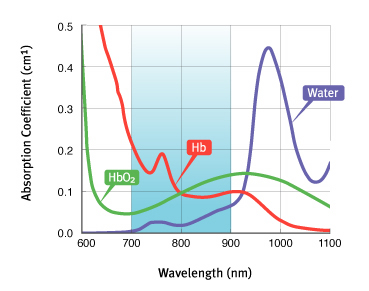
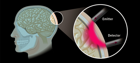
Imagent的工作原理是基于近红外光在皮质表面探测中的应用。跨越700 nm至900 nm波长范围的主要组织吸收体是含氧血红蛋白 (HbO2) 和脱氧血红蛋白 (Hb);在更小的尺度中,水、脂肪和细胞色素氧化酶都对光的部分吸收有帮助。在这个波长范围内,光在组织中的穿透深度相当显著。对于一个典型的头部组织 (皮肤/头皮,头骨和皮质层),其吸收系数为μa = 0.1 cm-1和散射系数为μs' = 8 cm-1,那么当探测器被放置在距光源4 cm处,最大光学穿透力可估算为1.5 cm左右。通过增加光源和探测器之间的距离,穿透深度可以被增加,尽管最后测量的信噪比会变差。
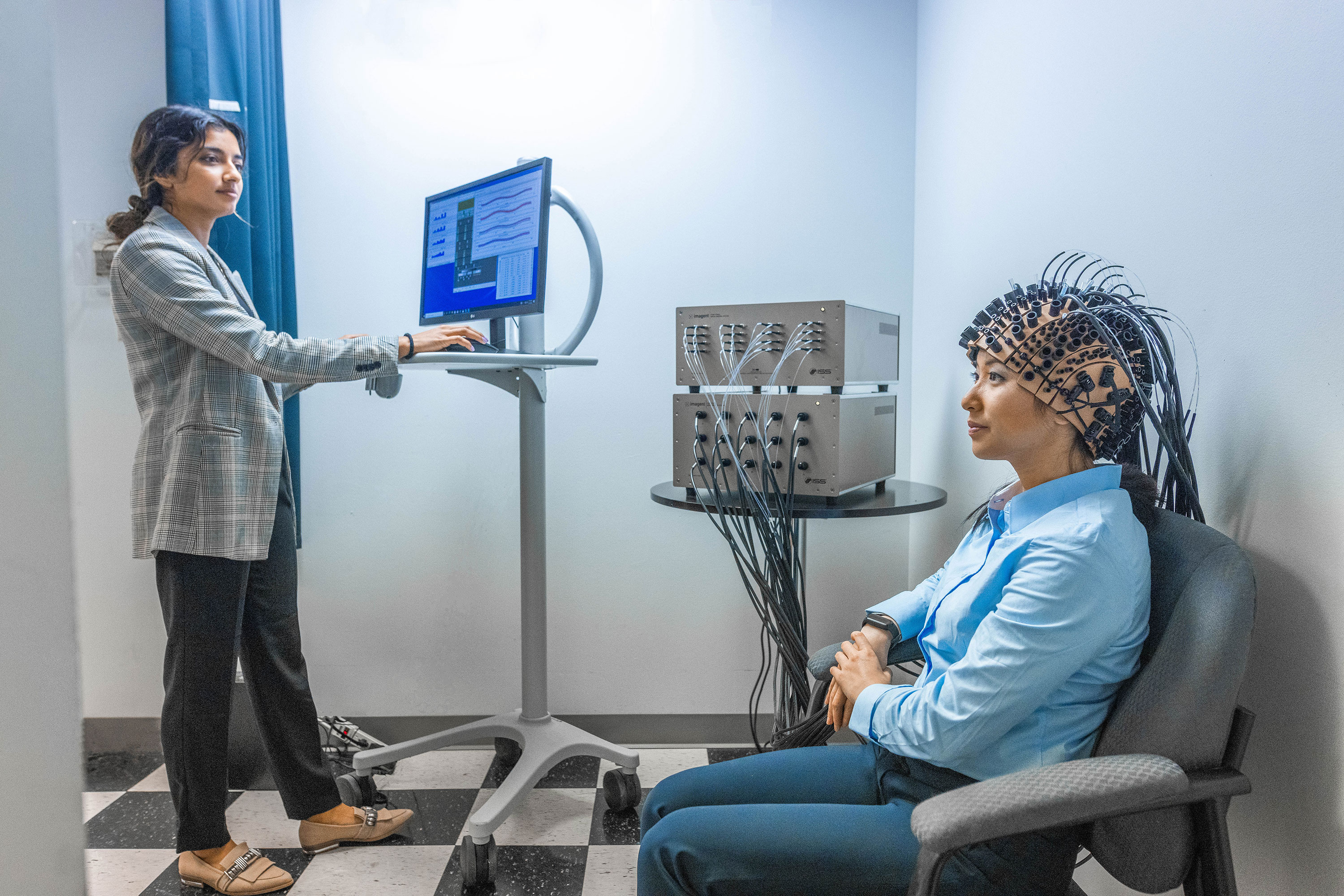
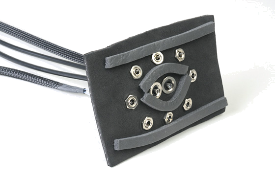
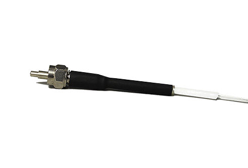
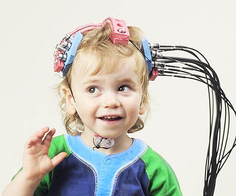
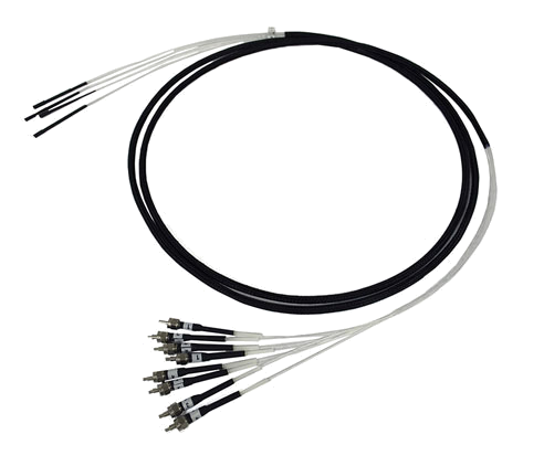
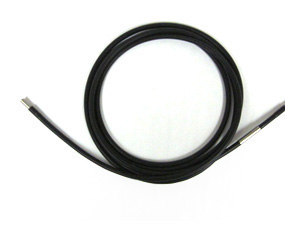
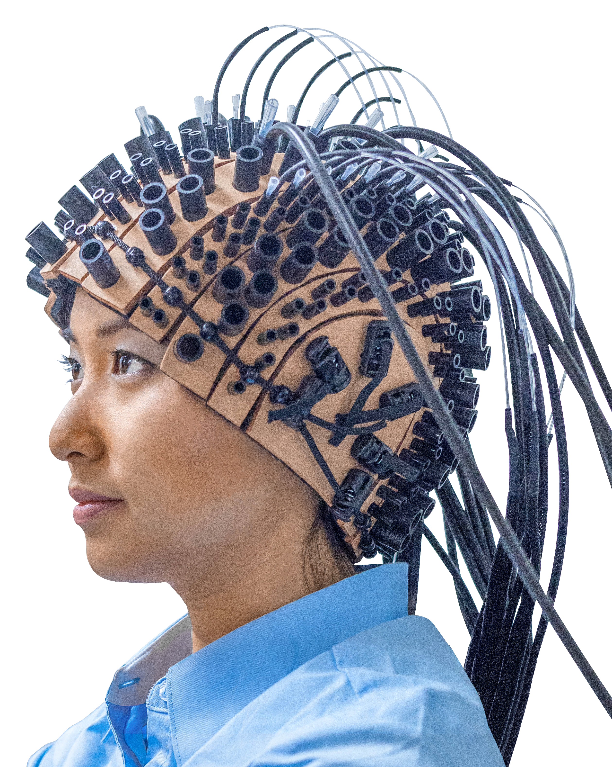
Imagent采用在690 nm和830 nm处发射的激光二极管。光被位于头部的光纤传递。近红外光进入组织后,虽然被微弱地吸收了,但它还是会被组织均匀地高度散射。一部分光离开组织,由收集光纤将其收集并带回该单元中的光探测器进行数据处理。光纤被一个头套所固定,大人和小孩都可以使用它;对于特定区域的研究,有垫片传感器可供使用。一个成年人的头部可以放置高达128根光纤和高达60个探测器束 (总共3,840个光学通道)。可以使用不同的激发图样 (蒙太奇) 和收集光纤。
Imagent采用频域技术,但在高频处 (100 MHz的量级) 对光源进行调制,并且测量了被探测信号的三个参数:强度、调制深度和穿过组织所需的时间 (相位延迟)。结合这三个测量量中的任意两个都可以被用作提供生理参数的变化,该选择取决于要测量的特定参数、减少生理噪声的需要和将要测量事件的时间尺度。
Product Specifications for Imagent
操作
- 方法:频域
- 调制频率 110 MHz
- 样本时间:最小15 ms
- 光学通道的数量:16至960
fNIRS测量参数
- O2Hb氧合血红蛋白的改变
- HHb脱氧血红蛋白的改变
- Hb血红蛋白总量的改变
EROS测量参数
- 信号强度
- 信号相位延迟
光源
- 纤维耦合激光二极管
- 波长:690 nm和830 nm
- 激光功率:平均10 mW
光探测器
- 光电倍增管
光极
- 激发的成对纤维,直径为400 µm
- 一系列纤维束,直径为3 mm
接口
- 可通过用于alairach脑坐标配准技术的Polhemus对接到FastTrack
前置放大器鉴频器
- 600 MHz带宽,TTL输出
计算机和操作系统
- 因特尔类型CPU,Windows 11操作系统
电源要求
- 通用电源输入:110 - 240 V, 250 W
Imagent的产品配件
产品资源
-
“On the functional role of temporal and frontal cortex activation in passive detection of auditory deviance.” Tse, C.-Y. & Penney, T.B. NeuroImage, 41(4), pp. 1462–1470, 2008, Jul. doi: 10.1016/j.neuroimage.2008.03.043.
-
“The role of visual and auditory temporal processing for Chinese children with developmental dyslexia.” Chung, K.K.H., Mcbride-Chang, C., Wong, S.W.L., Cheung, H., Penney, T.B. & Ho, C.S.-H. Annals of Dyslexia, 58(1), pp. 15–35, 2008, May. doi: 10.1007/s11881-008-0015-4.
-
“Optical imaging of temporal integration in human auditory cortex.” Sable, J.J., Low, K.A., Whalen, C.J., Maclin, E.L., Fabiani, M. & Gratton, G. European Journal of Neuroscience, 25(1), pp. 298–306, 2007, Jan. doi: 10.1111/j.1460-9568.2006.05255.x.
-
“Latent inhibition mediates N1 attenuation to repeating sounds.” Sable, J.J., Low, K.A., Maclin, E.L., Fabiani, M. & Gratton, G. Psychophysiology, 41(4), pp. 636–642, 2004, Jul. doi: 10.1111/j.1469-8986.2004.00192.x.
-
“RAPID COMMUNICATION Scalp-Recorded Optical Signals Make Sound Processing in the Auditory Cortex Visible?” Rinne, T., Gratton, G., Fabiani, M., Cowan, N., Maclin, E., Stinard, A., Sinkkonen, J., Alho, K. & Näätänen, R. NeuroImage, 10(5), pp. 620–624, 1999, Nov. doi: 10.1006/nimg.1999.0495.
-
“Functional near-infrared spectroscopy-based correlates of prefrontal cortical dynamics during a cognitive-motor executive adaptation task.” Gentili, R.J., Shewokis, P.A., Ayaz, H. & Contreras-Vidal, J.L. Frontiers in Human Neuroscience, 7, 2013. doi: 10.3389/fnhum.2013.00277.
-
“Number–Space Interactions in the Human Parietal Cortex: Enlightening the SNARC Effect with Functional Near-Infrared Spectroscopy.” Cutini, S., Scarpa, F., Scatturin, P., Dell'Acqua, R. & Zorzi, M. Cerebral Cortex, 24(2), pp. 444–451, 2012, Oct. doi: 10.1093/cercor/bhs321.
-
“Exploring the role of primary and supplementary motor areas in simple motor tasks with fNIRS.” Brigadoi, S., Cutini, S., Scarpa, F., Scatturin, P. & Dell'Acqua, R. Cognitive Processing, 13(S1), pp. 97–101, 2012, Jul. doi: 10.1007/s10339-012-0446-z.
-
“The cortical control of cycling exercise in stroke patients: An fNIRS study.” Lin, P.-Y., Chen, J.-J.J. & Lin, S.-I. Human Brain Mapping, 34(10), pp. 2381–2390, 2012, Mar. doi: 10.1002/hbm.22072.
-
“When in Doubt, Do it Both Ways: Brain Evidence of the Simultaneous Activation of Conflicting Motor Responses in a Spatial Stroop Task.” Desoto, M.C., Fabiani, M., Geary, D.C. & Gratton, G. Journal of Cognitive Neuroscience, 13(4), pp. 523–536, 2001, May. doi: 10.1162/08989290152001934.
-
“Rapid Changes of Optical Parameters in the Human Brain During a Tapping Task.” Gratton, G., Fabiani, M., Friedman, D., Franceschini, M.A., Fantini, S., Corballis, P. & Gratton, E. Journal of Cognitive Neuroscience, 7(4), pp. 446–456, 1995. doi: 10.1162/jocn.1995.7.4.446.
-
“Early childhood development of visual texture segregation in full-term and preterm children.” Sayeur, M.S., Vannasing, P., Lefrançois, M., Tremblay, E., Lepore, F., Lassonde, M., Mckerral, M. & Gallagher, A. Vision Research, 112, pp. 1–10, 2015, Jul. doi: 10.1016/j.visres.2015.04.013.
-
“Comparison of neuronal and hemodynamic measures of the brain response to visual stimulation: An optical imaging study.” Gratton, G., Goodman-Wood, M.R. & Fabiani, M. Human Brain Mapping, 13(1), pp. 13–25, 2001. doi: 10.1002/hbm.1021.
-
“Shades of gray matter: Noninvasive optical images of human brain reponses during visual stimulation.” Gratton, G., Corballis, P.M., Cho, E., Fabiani, M. & Hood, D.C. Psychophysiology, 32(5), pp. 505–509, 1995, Sep. doi: 10.1111/j.1469-8986.1995.tb02102.x.
-
“Distinct hemispheric specializations for native and non-native languages in one-day-old newborns identified by fNIRS.” Vannasing, P., Florea, O., González-Frankenberger, B., Tremblay, J., Paquette, N., Safi, D., Wallois, F., Lepore, F., Béland, R., Lassonde, M. & Gallagher, A. Neuropsychologia, 84, pp. 63–69, 2016, Apr. doi: 10.1016/j.neuropsychologia.2016.01.038.
-
“Early electrophysiological markers of atypical language processing in prematurely born infants.” Paquette, N., Vannasing, P., Tremblay, J., Lefebvre, F., Roy, M.-S., Mckerral, M., Lepore, F., Lassonde, M. & Gallagher, A. Neuropsychologia, 79, pp. 21–32, 2015, Dec. doi: 10.1016/j.neuropsychologia.2015.10.021.
-
“Developmental patterns of expressive language hemispheric lateralization in children, adolescents and adults using functional near-infrared spectroscopy.” Paquette, N., Lassonde, M., Vannasing, P., Tremblay, J., González-Frankenberger, B., Florea, O., Béland, R., Lepore, F. & Gallagher, A. Neuropsychologia, 68, pp. 117–125, 2015, Feb. doi: 10.1016/j.neuropsychologia.2015.01.007.
-
“Functional near-infrared spectroscopy for the assessment of overt reading.” Safi, D., Lassonde, M., Nguyen, D.K., Vannasing, P., Tremblay, J., Florea, O., Morin-Moncet, O., Lefrançois, M. & Béland, R. Brain and Behavior, 2(6), pp. 825–837, 2012, Oct. doi: 10.1002/brb3.100.
-
“Morphological Structure Awareness, Vocabulary, and Reading.” McBride-Chang, C., Hua, S., Ng, J.Y.L., Meng, X., & Penney, T.B. Vocabulary Acquisition: Implications for Reading Comprehension, 6, 2007, pp. 104 - 122.
-
“Speeded Naming and Dyslexia.” Penney, T.B., Wong, S., Ng, K.K., & McBride-Chang, C.A. Communicating Skills of Intention, 2007, pp. 75 - 90.
-
“Near-infrared Spectroscopy as an Alternative to the Wada Test for Language Mapping in Children, Adults and Special Populations.” Gallagher, A., Thériault, M., Maclin, E., Low, K., Gratton, G., Fabiani, M., Gagnon, L., Valois, K., Rouleau, I., Sauerwein, H.C., Carmant, L., Nguyen, D.K., Lortie, A., Lepore, F., Béland, R., & Lassonde, M. Epileptic Disord, 9(3), 2007, Sep.
-
“Poor readers of chinese respond slower than good readers in phonological, rapid naming, and interval timing tasks.” Penney, T.B., Leung, K.M., Chan, P.C., Meng, X. & Mcbride-Chang, C.A. Annals of Dyslexia, 55(1), pp. 9–27, 2005, Jun. doi: 10.1007/s11881-005-0002-y.
-
“Brain Responses to Segmentally and Tonally Induced Semantic Violations in Cantonese.” Schirmer, A., Tang, S.-L., Penney, T.B., Gunter, T.C. & Chen, H.-C. Journal of Cognitive Neuroscience, 17(1), pp. 1–12, 2005, Jan. doi: 10.1162/0898929052880057.
-
“Taking the pulse of aging: Mapping pulse pressure and elasticity in cerebral arteries with optical methods.” Fabiani, M., Low, K.A., Tan, C.-H., Zimmerman, B., Fletcher, M.A., Schneider-Garces, N., Maclin, E.L., Chiarelli, A.M., Sutton, B.P. & Gratton, G. Psychophysiology, 51(11), pp. 1072–1088, 2014, Aug. doi: 10.1111/psyp.12288.
-
“Contributions of Cognitive Neuroscience to the Understanding of Behavior and Aging.” Kramer, A.F., Fabiani, M. & Colcombe, S.J. Handbook of the Psychology of Aging, pp. 57–83, 2006. doi: 10.1016/b978-012101264-9/50007-0.
-
“Reduced Suppression or Labile Memory? Mechanisms of Inefficient Filtering of Irrelevant Information in Older Adults.” Fabiani, M., Low, K.A., Wee, E., Sable, J.J. & Gratton, G. Journal of Cognitive Neuroscience, 18(4), pp. 637–650, 2006, Apr. doi: 10.1162/jocn.2006.18.4.637.
-
“Multiple Electrophysiological Indices of Distinctiveness.” Fabiani, M. Distinctiveness and Memory, pp. 338–360, 2006, Apr. doi: 10.1093/acprof:oso/9780195169669.003.0015.
-
“Electrophysiological and Optical Measures of Cognitive Aging.” Fabiani, M. & Gratton, G. , pp. 85–106, 2004, Dec. doi: 10.1093/acprof:oso/9780195156744.003.0004.
-
“Sensory ERPs predict differences in working memory span and fluid intelligence.” Brumback, C.R., Low, K.A., Gratton, G. & Fabiani, M. NeuroReport, 15(2), pp. 373–376, 2004, Feb. doi: 10.1097/00001756-200402090-00032.
-
“Recruitment of the left precentral gyrus in reading epilepsy: A multimodal neuroimaging study.” Safi, D., Béland, R., Nguyen, D.K., Pouliot, P., Mohamed, I.S., Vannasing, P., Tremblay, J., Lassonde, M. & Gallagher, A. Epilepsy & Behavior Case Reports, 5, pp. 19–22, 2016. doi: 10.1016/j.ebcr.2016.01.003.
-
“Potential brain language reorganization in a boy with refractory epilepsy; an fNIRS–EEG and fMRI comparison.” Vannasing, P., Cornaggia, I., Vanasse, C., Tremblay, J., Diadori, P., Perreault, S., Lassonde, M. & Gallagher, A. Epilepsy & Behavior Case Reports, 5, pp. 34–37, 2016. doi: 10.1016/j.ebcr.2016.01.006.
-
“Using patient-specific hemodynamic response function in epileptic spike analysis of human epilepsy: a study based on EEG–fNIRS.” Peng, K., Nguyen, D.K., Vannasing, P., Tremblay, J., Lesage, F. & Pouliot, P. NeuroImage, 126, pp. 239–255, 2016, Feb. doi: 10.1016/j.neuroimage.2015.11.045.
-
“Hemodynamic changes during posterior epilepsies: An EEG-fNIRS study.” Pouliot, P., Tran, T.P.Y., Birca, V., Vannasing, P., Tremblay, J., Lassonde, M. & Nguyen, D.K. Epilepsy Research, 108(5), pp. 883–890, 2014, Jul. doi: 10.1016/j.eplepsyres.2014.03.007.
-
“fNIRS-EEG study of focal interictal epileptiform discharges.” Peng, K., Nguyen, D.K., Tayah, T., Vannasing, P., Tremblay, J., Sawan, M., Lassonde, M., Lesage, F. & Pouliot, P. Epilepsy Research, 108(3), pp. 491–505, 2014, Mar. doi: 10.1016/j.eplepsyres.2013.12.011.
-
“Noninvasive continuous functional near-infrared spectroscopy combined with electroencephalography recording of frontal lobe seizures.” Nguyen, D.K., Tremblay, J., Pouliot, P., Vannasing, P., Florea, O., Carmant, L., Lepore, F., Sawan, M., Lesage, F. & Lassonde, M. Epilepsia, 54(2), pp. 331–340, 2012, Nov. doi: 10.1111/epi.12011.
-
“Nonlinear hemodynamic responses in human epilepsy: A multimodal analysis with fNIRS-EEG and fMRI-EEG.” Pouliot, P., Tremblay, J., Robert, M., Vannasing, P., Lepore, F., Lassonde, M., Sawan, M., Nguyen, D.K. & Lesage, F. Journal of Neuroscience Methods, 204(2), pp. 326–340, 2012, Mar. doi: 10.1016/j.jneumeth.2011.11.016.
-
“Perception of Caucasian and African faces in 5- to 9-month-old Caucasian infants: A functional near-infrared spectroscopy study.” Timeo, S., Brigadoi, S. & Farroni, T. Neuropsychologia, 126, pp. 3–9, 2019, Mar. doi: 10.1016/j.neuropsychologia.2017.09.011.
-
“Infant cortex responds to other humans from shortly after birth.” Farroni, T., Chiarelli, A.M., Lloyd-Fox, S., Massaccesi, S., Merla, A., Gangi, V.D., Mattarello, T., Faraguna, D. & Johnson, M.H. Scientific Reports, 3(1), 2013, Oct. doi: 10.1038/srep02851.
-
“Coupled oxygenation oscillation measured by NIRS and intermittent cerebral activation on EEG in premature infants.” Roche-Labarbe, N., Wallois, F., Ponchel, E., Kongolo, G. & Grebe, R. NeuroImage, 36(3), pp. 718–727, 2007, Jul. doi: 10.1016/j.neuroimage.2007.04.002.
-
“Measurement of brain activity by near-infrared light.” Gratton, E., Toronov, V., Wolf, U., Wolf, M. & Webb, A. Journal of Biomedical Optics, 10(1), p. 011008, 2005. doi: 10.1117/1.1854673.
-
“Noninvasive determination of the optical properties of adult brain: near-infrared spectroscopy approach.” Choi, J., Wolf, M., Toronov, V., Wolf, U., Polzonetti, C., Hueber, D., Safonova, L.P., Gupta, R., Michalos, A., Mantulin, W. & Gratton, E. Journal of Biomedical Optics, 9(1), p. 221, 2004. doi: 10.1117/1.1628242.
-
“Absolute Frequency-Domain Pulse Oximetry of the Brain: Methodology and Measurements.” Wolf, M., Franceschini, M.A., Paunescu, L.A., Toronov, V., Michalos, A., Wolf, U., Gratton, E. & Fantini, S. Oxygen Transport to Tissue, 24, pp. 61–73, 2003. doi: 10.1007/978-1-4615-0075-9_7.
-
“Fast cerebral functional signal in the 100-ms range detected in the visual cortex by frequency-domain near-infrared spectrophotometry.” Wolf, M., Wolf, U., Choi, J.H., Toronov, V., Paunescu, L.A., Michalos, A. & Gratton, E. Psychophysiology, 40(4), pp. 521–528, 2003, Jul. doi: 10.1111/1469-8986.00054.
-
“Different Time Evolution of Oxyhemoglobin and Deoxyhemoglobin Concentration Changes in the Visual and Motor Cortices during Functional Stimulation: A Near-Infrared Spectroscopy Study.” Wolf, M., Wolf, U., Toronov, V., Michalos, A., Paunescu, L., Choi, J.H. & Gratton, E. NeuroImage, 16(3), pp. 704–712, 2002, Jul. doi: 10.1006/nimg.2002.1128.
-
“Near-infrared study of fluctuations in cerebral hemodynamics during rest and motor stimulation: Temporal analysis and spatial mapping.” Toronov, V., Franceschini, M.A., Filiaci, M., Fantini, S., Wolf, M., Michalos, A. & Gratton, E. Medical Physics, 27(4), pp. 801–815, 2000, Apr. doi: 10.1118/1.598943.
-
“Cerebral hemodynamics measured by near-infrared spectroscopy at rest and during motor activation.” Angela, M., Sergio, F., Sergio, F., Vlad, T., Mattia, F. & Enrico, G. Proceedings of the Optical Society of America In Vivo Optical Imaging Workshop: Washington, pp. 78 - 80, 2000, Jan.
-
“On-line optical imaging of the human brain with 160-ms temporal resolution.” Franceschini, M.A., Toronov, V., Filiaci, M.E., Gratton, E. & Fantini, S. Optics Express, 6(3), p. 49, 2000, Jan. doi: 10.1364/oe.6.000049.
-
“Shades of gray matter: Noninvasive optical images of human brain reponses during visual stimulation.” Gratton, G., Corballis, P.M., Cho, E., Fabiani, M. & Hood, D.C. Psychophysiology, 32(5), pp. 505–509, 1995, Sep. doi: 10.1111/j.1469-8986.1995.tb02102.x.
-
“Dynamic filtering improves attentional state prediction with fNIRS.” Harrivel, A.R., Weissman, D.H., Noll, D.C., Huppert, T. & Peltier, S.J. Biomedical Optics Express, 7(3), p. 979, 2016, Feb. doi: 10.1364/boe.7.000979.
-
“Detection of Attentional State in Long-Distance Driving Settings Using Functional Near-Infrared Spectroscopy.” Matthew, T., Miles, A., V., S., Michael, C. & Mary, C. 95th Annual Transportation Research Board Meeting, 2016, Jan.
-
“Investigating Mental Workload Changes in a Long Duration Supervisory Control Task.” Boyer, M., Cummings, M., Spence, L.B. & Solovey, E.T. Interacting with Computers, 27(5), pp. 512–520, 2015, May. doi: 10.1093/iwc/iwv012.
-
“Monitoring attentional state with fNIRS.” Harrivel, A.R., Weissman, D.H., Noll, D.C. & Peltier, S.J. Frontiers in Human Neuroscience, 7, 2013. doi: 10.3389/fnhum.2013.00861.
-
“Learn Piano with BACh.” Yuksel, B.F., Oleson, K.B., Harrison, L., Peck, E.M., Afergan, D., Chang, R. & Jacob, R.J. Proceedings of the 2016 CHI Conference on Human Factors in Computing Systems, 2016, May. doi: 10.1145/2858036.2858388.
-
“Braahms: a novel adaptive musical interface based on users' cognitive state.” Yuksel, B.F., Afergan, D., Peck, E.M., Griffin, G., Harrison, L., Chen, N.W.B., Chang, R., & Jacob, R.J.K. NIME, pp. 136–139, 2015.
-
“A hybrid NIRS-EEG system for self-paced brain computer interface with online motor imagery.” Koo, B., Lee, H.-G., Nam, Y., Kang, H., Koh, C.S., Shin, H.-C. & Choi, S. Journal of Neuroscience Methods, 244, pp. 26–32, 2015, Apr. doi: 10.1016/j.jneumeth.2014.04.016.
-
“Brain-based target expansion.” Afergan, D., Shibata, T., Hincks, S.W., Peck, E.M., Yuksel, B.F., Chang, R. & Jacob, R.J. Proceedings of the 27th annual ACM symposium on User interface software and technology, 2014, Oct. doi: 10.1145/2642918.2647414.
-
“Temporal decoupling of oxy- and deoxy-hemoglobin hemodynamic responses detected by functional near-infrared spectroscopy (fNIRS).” Tam, N.D. & Zouridakis, G. Journal of Biomedical Engineering and Medical Imaging, 1(2), pp. 18–28, 2014, Apr. doi: 10.14738/jbemi.12.146.
-
“Using fNIRS brain sensing to evaluate information visualization interfaces.” Peck, E.M.M., Yuksel, B.F., Ottley, A., Jacob, R.J. & Chang, R. Proceedings of the SIGCHI Conference on Human Factors in Computing Systems, 2013, Apr. doi: 10.1145/2470654.2470723.
-
“Classification of prefrontal activity due to mental arithmetic and music imagery using hidden Markov models and frequency domain near-infrared spectroscopy.” Power, S.D., Falk, T.H. & Chau, T. Journal of Neural Engineering, 7(2), p. 026002, 2010, Feb. doi: 10.1088/1741-2560/7/2/026002.
-
“Single-trial classification of NIRS signals during emotional induction tasks: towards a corporeal machine interface.” Tai, K. & Chau, T. Journal of NeuroEngineering and Rehabilitation, 6(1), 2009, Nov. doi: 10.1186/1743-0003-6-39.
-
“Taking NIRS-BCIs Outside the Lab: Towards Achieving Robustness Against Environment Noise.” Falk, T.H., Guirgis, M., Power, S. & Chau, T.T. IEEE Transactions on Neural Systems and Rehabilitation Engineering, 19(2), pp. 136–146, 2011, Apr. doi: 10.1109/tnsre.2010.2078516.
-
“A new method based on ICBM152 head surface for probe placement in multichannel fNIRS.” Cutini, S., Scatturin, P. & Zorzi, M. NeuroImage, 54(2), pp. 919–927, 2011, Jan. doi: 10.1016/j.neuroimage.2010.09.030.
-
“Bayesian filtering of human brain hemodynamic activity elicited by visual short-term maintenance recorded through functional near-infrared spectroscopy (fNIRS).” Scarpa, F., Cutini, S., Scatturin, P., Dell'Acqua, R. & Sparacino, G. Optics Express, 18(25), p. 26550, 2010, Dec. doi: 10.1364/oe.18.026550.
-
“Validation of a method for coregistering scalp recording locations with 3D structural MR images.” Whalen, C., Maclin, E.L., Fabiani, M. & Gratton, G. Human Brain Mapping, 29(11), pp. 1288–1301, 2008, Nov. doi: 10.1002/hbm.20465.
-
“Level-set algorithm for the reconstruction of functional activation in near-infrared spectroscopic imaging.” Jacob, M., Bresler, Y., Toronov, V., Zhang, X. & Webb, A. Journal of Biomedical Optics, 11(6), p. 064029, 2006. doi: 10.1117/1.2400595.
-
“Signal and image processing techniques for functional near-infrared imaging of the human brain.” Toronov, V.Y., Zhang, X., Fabiani, M., Gratton, G. & Webb, A.G. SPIE Proceedings, 2005, Mar. doi: 10.1117/12.593345.
-
“Optimization of the frequency-domain instrument for the near-infrared spectro-imaging of the human brain.” Toronov, V.Y., Di Amico, E., Hueber, D., Gratton, E., Webb, A.G. & Barbieri, B.B. SPIE Proceedings, 2004, Jul. doi: 10.1117/12.530254.
-
“Optimization of the signal-to-noise ratio of frequency-domain instrumentation for near-infrared spectro-imaging of the human brain.” Toronov, V., D'Amico, E., Hueber, D., Gratton, E., Barbieri, B. & Webb, A. Optics Express, 11(21), p. 2717, 2003, Oct. doi: 10.1364/oe.11.002717.
-
“Frequency-Domain Techniques for Cerebral and Functional Near-Infrared Spectroscopy.” Fantini, S. & Sassaroli, A. Frontiers in Neuroscience, 14, 2020, Apr. doi: 10.3389/fnins.2020.00300.
-
“A brief review on the history of human functional near-infrared spectroscopy (fNIRS) development and fields of application.” Ferrari, M. & Quaresima, V. NeuroImage, 63(2), pp. 921–935, 2012, Nov. doi: 10.1016/j.neuroimage.2012.03.049.
-
“Detection of event-related hemodynamic response to neuroactivation by dynamic modeling of brain activity.” Aqil, M., Hong, K.-S., Jeong, M.-Y. & Ge, S.S. NeuroImage, 63(1), pp. 553–568, 2012, Oct. doi: 10.1016/j.neuroimage.2012.07.006.
-
“Cortical brain imaging by adaptive filtering of NIRS signals.” Aqil, M., Hong, K.-S., Jeong, M.-Y. & Ge, S.S. Neuroscience Letters, 514(1), pp. 35–41, 2012, Apr. doi: 10.1016/j.neulet.2012.02.048.
-
“Functional near Infrared Optical Imaging in Cognitive Neuroscience: An Introductory Review.” Cutini, S., Moro, S.B. & Bisconti, S. Journal of Near Infrared Spectroscopy, 20(1), pp. 75–92, 2012, Jan. doi: 10.1255/jnirs.969.
-
“Near Infrared Brain and Muscle Oximetry: From the Discovery to Current Applications.” Ferrari, M. & Quaresima, V. Journal of Near Infrared Spectroscopy, 20(1), pp. 1–14, 2012, Jan. doi: 10.1255/jnirs.973.
-
“The influence of posterior parietal cortex on extrastriate visual activity: A concurrent TMS and fast optical imaging study.” Parks, N.A., Mazzi, C., Tapia, E., Savazzi, S., Fabiani, M., Gratton, G. & Beck, D.M. Neuropsychologia, 78, pp. 153–158, 2015, Nov. doi: 10.1016/j.neuropsychologia.2015.10.002.
-
“Preattentive change detection using the event-related optical signal.” Tse, C.-Y. & Penney, T.B. IEEE Engineering in Medicine and Biology Magazine, 26(4), pp. 52–58, 2007, Jul. doi: 10.1109/memb.2007.384096.
-
“Event-related optical imaging reveals the temporal dynamics of right temporal and frontal cortex activation in pre-attentive change detection.” Tse, C.-Y., Tien, K.-R. & Penney, T.B. NeuroImage, 29(1), pp. 314–320, 2006, Jan. doi: 10.1016/j.neuroimage.2005.07.013.
-
“Fast optical signal in visual cortex: Improving detection by General Linear Convolution Model.” Chiarelli, A.M., Vacri, A.D., Romani, G.L. & Merla, A. NeuroImage, 66, pp. 194–202, 2013, Feb. doi: 10.1016/j.neuroimage.2012.10.047.
-
“Time course of activation of human occipital cortex measured with the event-related optical signal (EROS).” Gratton, G., Low, K.A., Maclin, E.L., Brumback, C.R., Gordon, B. & Fabiani, M. Biomedical Optics, 2006. doi: 10.1364/bio.2006.md4.
-
“Fronto-occipital mismatch responses in pre-attentive detection of visual changes: Implication on a generic brain network underlying Mismatch Negativity (MMN).” Tse, C.-Y., Shum, Y.-H., Xiao, X.-Z. & Wang, Y. NeuroImage, 244, p. 118633, 2021, Dec. doi: 10.1016/j.neuroimage.2021.118633.
-
“Functional connectivity of the frontotemporal network in preattentive detection of abstract changes: Perturbs and observes with transcranial magnetic stimulation and event-related optical signal.” Xiao, X.-Z., Shum, Y.-H., Lui, T.K.-Y., Wang, Y., Cheung, A.T.-C., Chu, W.C.W., Neggers, S.F.W., Chan, S.S.-M. & Tse, C.-Y. Human Brain Mapping, 41(11), pp. 2883–2897, 2020, Mar. doi: 10.1002/hbm.24984.
-
“Time course of activation of human occipital cortex measured with the event-related optical signal (EROS).” Gratton, G., Low, K.A., Maclin, E.L., Brumback, C.R., Gordon, B. & Fabiani, M. Biomedical Optics, 2006. doi: 10.1364/bio.2006.md4.
-
“Fast optical imaging of frontal cortex during active and passive oddball tasks.” Low, K.A., Leaver, E., Kramer, A.F., Fabiani, M. & Gratton, G. Psychophysiology, 43(2), pp. 127–136, 2006, Mar. doi: 10.1111/j.1469-8986.2006.00390.x.
-
“Dynamic brain imaging: Event-related optical signal (EROS) measures of the time course and localization of cognitive-related activity.” Gratton, G. & Fabiani, M. Psychonomic Bulletin & Review, 5(4), pp. 535–563, 1998, Dec. doi: 10.3758/bf03208834.
-
“Cortical Dynamics of Semantic Processing during Sentence Comprehension: Evidence from Event-Related Optical Signals.” Huang, J., Wang, S., Jia, S., Mo, D. & Chen, H.-C. PLoS ONE, 8(8), p. e70671, 2013, Aug. doi: 10.1371/journal.pone.0070671.
-
“Imaging cortical dynamics of language processing with the event-related optical signal.” Tse, C.-Y., Lee, C.-L., Sullivan, J., Garnsey, S.M., Dell, G.S., Fabiani, M. & Gratton, G. Proceedings of the National Academy of Sciences, 104(43), pp. 17157–17162, 2007, Oct. doi: 10.1073/pnas.0707901104.
-
“
Proof-of-concept evidence for trimodal simultaneous investigation of human brain function.” Moore, M., Maclin, E.L., Iordan, A.D., Katsumi, Y., Larsen, R.J., Bagshaw, A.P., Mayhew, S., Shafer, A.T., Sutton, B.P., Fabiani, M., Gratton, G. & Dolcos, F. Human Brain Mapping, 42(13), pp. 4102–4121, 2021, Jun. doi: 10.1002/hbm.25541. -
“Establishing the functional connectivity of the frontotemporal network in pre-attentive change detection with Transcranial Magnetic Stimulation and event-related optical signal.” Tse, C.-Y., Yip, L.-Y., Lui, T.K.-Y., Xiao, X.-Z., Wang, Y., Chu, W.C.W., Parks, N.A., Chan, S.S.-M. & Neggers, S.F.W. NeuroImage, 179, pp. 403–413, 2018, Oct. doi: 10.1016/j.neuroimage.2018.06.053.
-
“Validation of a method for coregistering scalp recording locations with 3D structural MR images.” Whalen, C., Maclin, E.L., Fabiani, M. & Gratton, G. Human Brain Mapping, 29(11), pp. 1288–1301, 2008, Nov. doi: 10.1002/hbm.20465.
-
“Improving the signal-to-noise ratio of event-related optical signals.” Maclin, E.L., Low, K.A., Fabiani, M. & Gratton, G. IEEE Engineering in Medicine and Biology Magazine, 26(4), pp. 47–51, 2007, Jul. doi: 10.1109/memb.2007.384095.
-
“Effects of measurement method, wavelength, and source-detector distance on the fast optical signal.” Gratton, G., Brumback, C.R., Gordon, B.A., Pearson, M.A., Low, K.A. & Fabiani, M. NeuroImage, 32(4), pp. 1576–1590, 2006, Oct. doi: 10.1016/j.neuroimage.2006.05.030.
-
“Lagged covariance structure models for studying functional connectivity in the brain.” Rykhlevskaia, E., Fabiani, M. & Gratton, G. NeuroImage, 30(4), pp. 1203–1218, 2006, May. doi: 10.1016/j.neuroimage.2005.11.019.
-
“Optimum filtering for EROS measurements.” Maclin, E.L., Gratton, G. & Fabiani, M. Psychophysiology, 40(4), pp. 542–547, 2003, Jul. doi: 10.1111/1469-8986.00056.
-
“The event-related optical signal (EROS) in visual cortex: Replicability, consistency, localization, and resolution.” Gratton, G. & Fabiani, M. Psychophysiology, 40(4), pp. 561–571, 2003, Jul. doi: 10.1111/1469-8986.00058.
-
“Toward Noninvasive 3-D Imaging of the Time Course of Cortical Activity: Investigation of the Depth of the Event-Related Optical Signal.” Gratton, G., Sarno, A., Maclin, E., Corballis, P.M. & Fabiani, M. NeuroImage, 11(5), pp. 491–504, 2000, May. doi: 10.1006/nimg.2000.0565.
-
“Bootstrap assessment of the reliability of maxima in surface maps of brain activity of individual subjects derived with electrophysiological and optical methods.” Fabiani, M., Gratton, G., Corballis, P.M., Cheng, J. & Friedman, D. {Behavior Research Methods, Instruments, 30(1), pp. 78–86, 1998, Mar. doi: 10.3758/bf03209418.
-
“Principles, Methods, and Experimental Results.” Gratton, G., & Fabiani, M. In: Frostig, R.D., editor. In Vivo Optical Imaging of Brain Function. 2nd edition. Boca Raton (FL): CRC Press/Taylor & Francis; 2009. Chapter 15.
-
“Optical imaging of the intact human brain [Guest Editorial].” Fabiani, M., Schmorrow, D.D. & Gratton, G. IEEE Engineering in Medicine and Biology Magazine, 26(4), pp. 14–16, 2007, Jul. doi: 10.1109/memb.2007.384090.
-
“Event-Related Brain Potentials.” Fabiani, M., Gratton, G., & Federmeier, K.D. In Cacioppo, J., Tassinary, L., & Berntson, G. (Eds.), Handbook of Psychophysiology, pp. 85 - 119, 2007, Jun. doi: 10.1017/cbo9780511546396.
-
“Optical Imaging of Brain Function.” Gratton, G. & Fabiani, M. Neuroergonomics, pp. 65–81, 2006, Nov. doi: 10.1093/acprof:oso/9780195177619.003.0005.
-
“Lagged covariance structure models for studying functional connectivity in the brain.” Rykhlevskaia, E., Fabiani, M. & Gratton, G. NeuroImage, 30(4), pp. 1203–1218, 2006, May. doi: 10.1016/j.neuroimage.2005.11.019.
-
“Fast cerebral functional signal in the 100-ms range detected in the visual cortex by frequency-domain near-infrared spectrophotometry.” Wolf, M., Wolf, U., Choi, J.H., Toronov, V., Paunescu, L.A., Michalos, A. & Gratton, E. Psychophysiology, 40(4), pp. 521–528, 2003, Jul. doi: 10.1111/1469-8986.00054.
-
“Seeing right through you: Applications of optical imaging to the study of the human brain.” Gratton, G., Fabiani, M., Elbert, T. & Rockstroh, B. Psychophysiology, 40(4), pp. 487–491, 2003, Jul. doi: 10.1111/1469-8986.00050.
-
“The event-related optical signal: a new tool for studying brain function.” Gratton, G. & Fabiani, M. International Journal of Psychophysiology, 42(2), pp. 109–121, 2001, Oct. doi: 10.1016/s0167-8760(01)00161-1.
-
“Shedding light on brain function: the event-related optical signal.” Gratton, G. & Fabiani, M. Trends in Cognitive Sciences, 5(8), pp. 357–363, 2001, Aug. doi: 10.1016/s1364-6613(00)01701-0.
-
“What Triggers the Interictal Epileptic Spike? A Multimodal Multiscale Analysis of the Dynamic of Synaptic and Non-synaptic Neuronal and Vascular Compartments Using Electrical and Optical Measurements.” Arnal-Real, C., Mahmoudzadeh, M., Manoochehri, M., Nourhashemi, M. & Wallois, F. Frontiers in Neurology, 12, 2021, Feb. doi: 10.3389/fneur.2021.596926.
-
“Cortical hemodynamic mapping of subthalamic nucleus deep brain stimulation in Parkinsonian patients, using high-density functional near-infrared spectroscopy.” Mahmoudzadeh, M., Wallois, F., Tir, M., Krystkowiak, P. & Lefranc, M. PLOS ONE, 16(1), p. e0245188, 2021, Jan. doi: 10.1371/journal.pone.0245188.
