


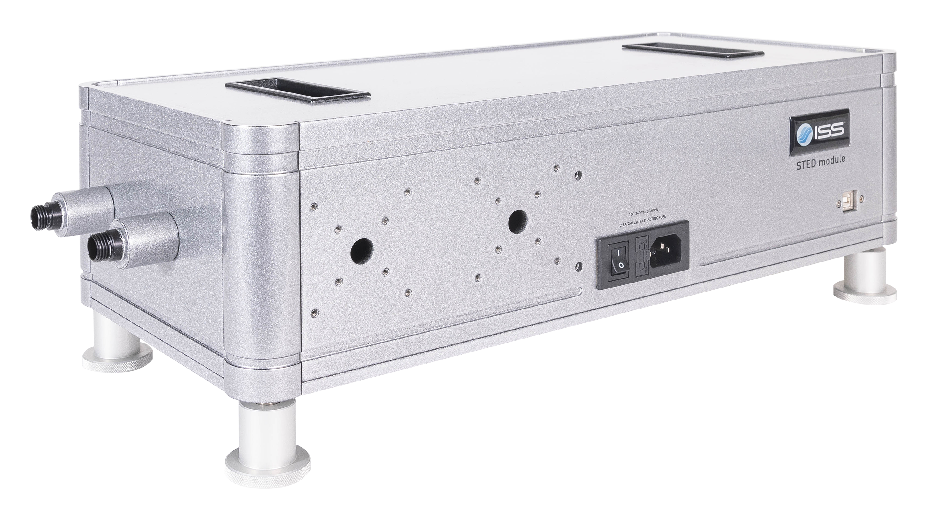
STED Module

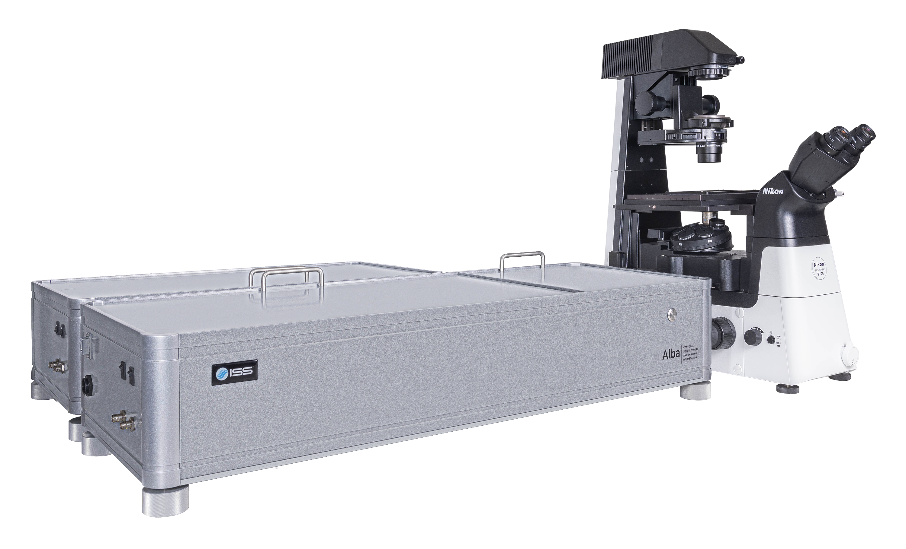
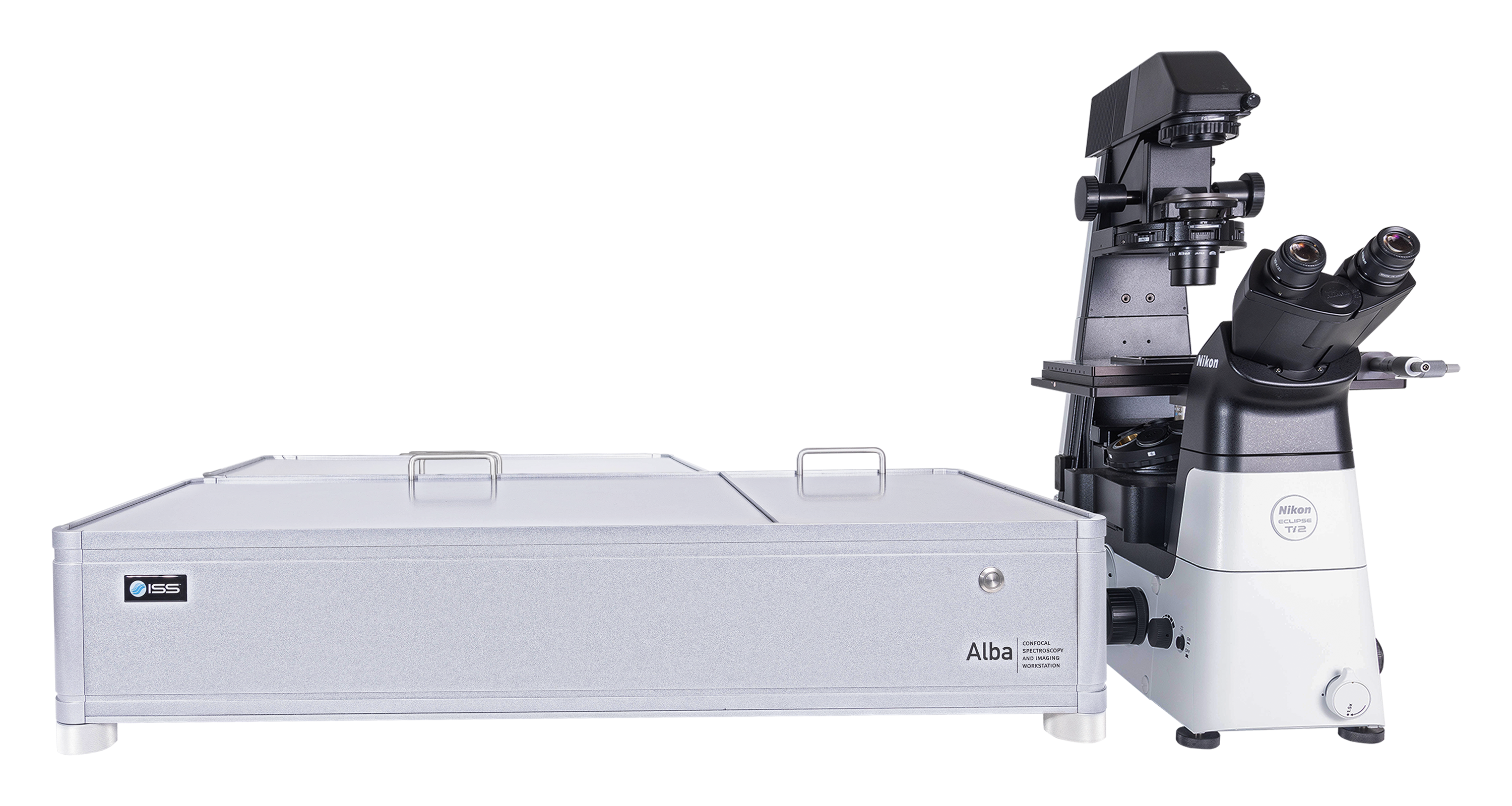
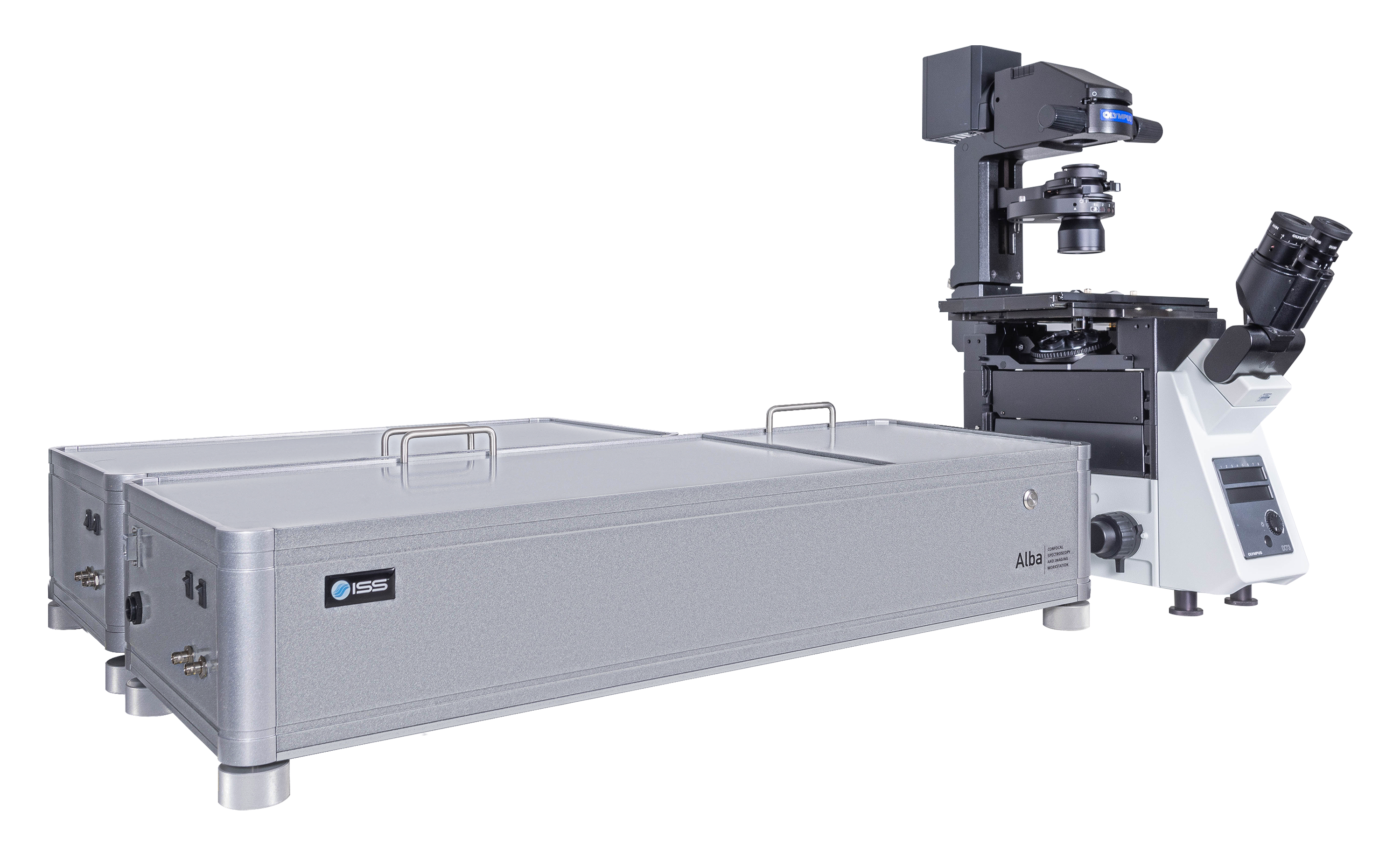
Alba v5 is a laser scanning microscope that incorporates several measurement modalities for experimental quantitative biology and material science applications requiring single molecule detection sensitivity. Capable of acquisition from the violet to the near infrared region, it features two independent laser entry ports. Alba v5 is powered by VistaVision, the comprehensive software package for instrument control, image acquisition and processing.

FLIM FRET in Live Cells

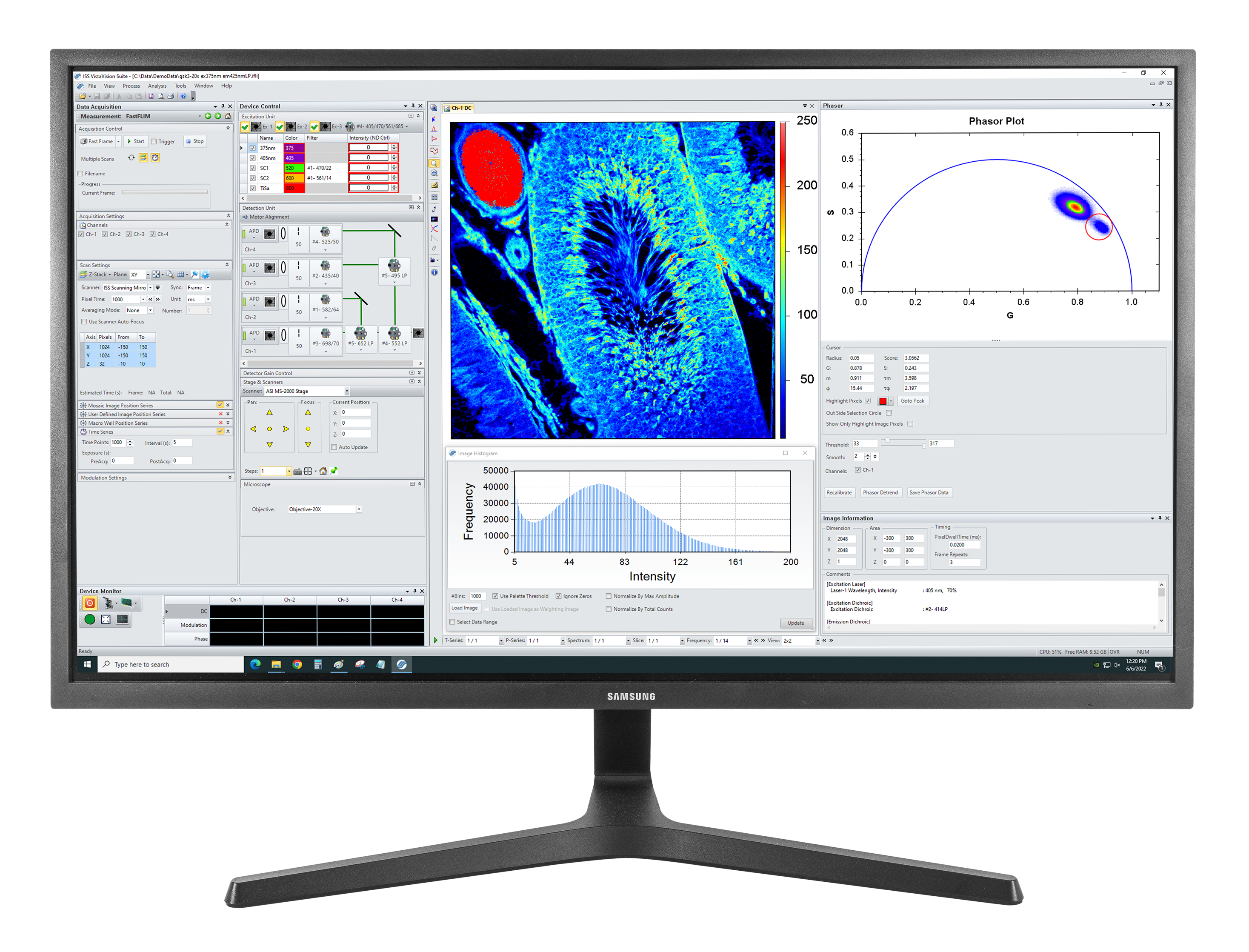
Tissue Imaging

Single Molecule Studies & Detection of Single Emitters by Antibunching

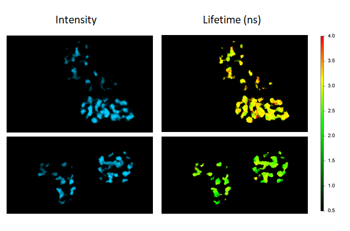
Multiplex Imaging by Time Resolved Unmixing Using the Phasor Plot

Multi-modality Imaging
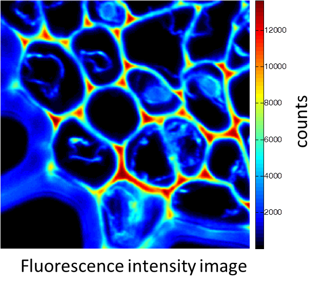
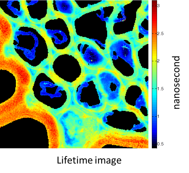
Rhizome cross-section of Convallaria (Lily of the Valley). Images were taken simultaneously using an Alba equipped with a multiphoton laser emitting at 800 nm. The standard fluorescence intensity image (first), the fluorescence lifetime image (second) and the second-harmonic generation (SHG) image (third). All images are 256 × 256 pixels, 40 × 40 µm2.
(courtesy of Dr. Zhang, Beckman Institute, Urbana, IL)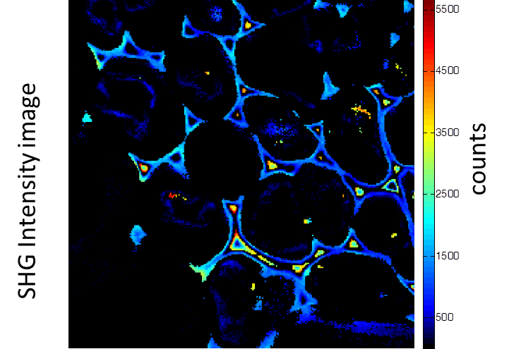
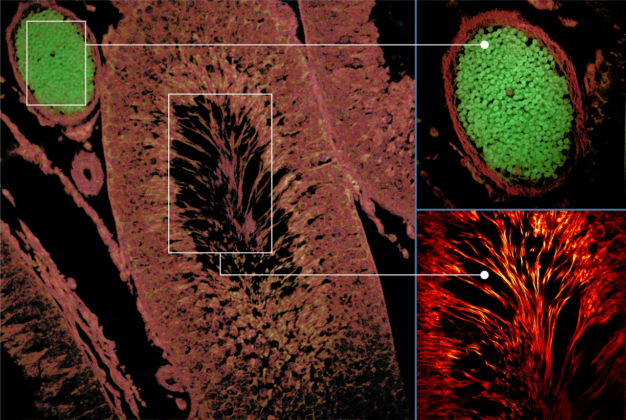
Autofluorescence tissue image: Excitation: 375 nm, Emission: 425 nm, Objective: 20X NA0.75
(courtesy of Dr. Aneesh Alex, GSK)
FLIM-FRET maps the FRET efficiencies of Cebp/α proteins co-expressing Cerulean and Venus in live cells to localize their dimerization in the cell nuclei (donor-only control: cells expressing Cebp/α-Cerulean only; FRET sample: cells co-expressing Cebp/α-Cerulean and Cebp/α-Venus).
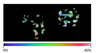
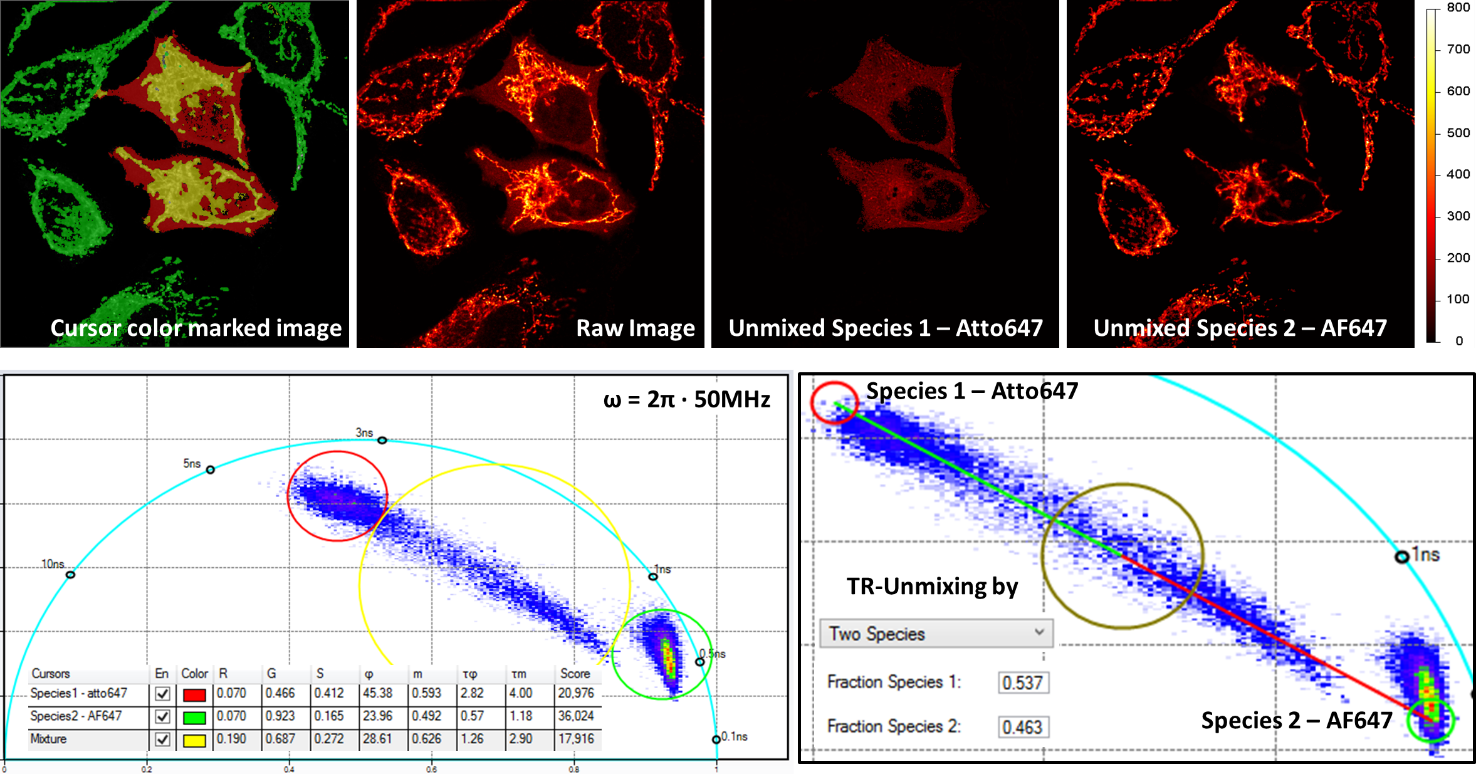
Multiplex imaging by time-resolved fluorescence requires using one excitation wavelength, and data is acquired on one detection channel. The figure shows the multiplexing of two labels, mitochondria labeled with Alexa Fluor 647 (AF647) and microtubule labeled with Atto 647 (Atto647) in the same cells. The raw image is acquired with excitation at 640 nm and detection in the same emission channel. Using the analysis routine in the phasor plot, the two structures are separated.
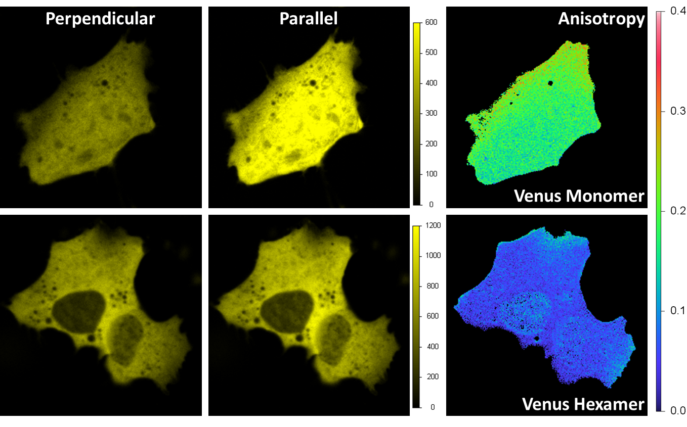
Venus was expressed in a HeLa cell. Excitation is 514 nm and emission is through a band-pass filter 545/35 nm. A beam-splitter polarizer in the emission splits the fluorescence beam into a perpendicular and parallel orientation beams, with each beam detected by one acquisition channel. The anisotropy image is calculated at each pixel. For the monomer (upper right) the average value of the anisotropy is around 0.2 indicating a prevalent orientation along the parallel axis; for the hexamer is less than 0.1 due to the local homo FRET between different Venus fluorescent proteins on the hexamer structure.
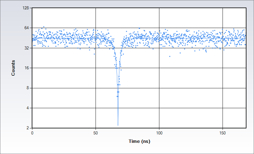
Antibunching data acquired on a solution of Rhodamine 110 in water with excitation at 488 nm. Measurements are acquired by splitting the signal in two beams following the classic Hanbury-Brown-Twiss set up and sending each of them to a separate detector of the Alba: the acquisition electronics provides a histogram of the time difference between the arrival time of the photons at the detectors. The histogram displays a dip at the coincidence point; the depth of the dip depends on the number of independent emitters in the observation volume while the shape depends on the excited state lifetime.

A peristaltic pump supplies the stage with a solution for keeping the sample conditions (temperature, pH, etc...) stable.

When using water objectives for prolonged measurements, it prevents the liquid drying up.

It keeps the focus position of the objective for hours using an active feedback to counter drifts.

Stage top incubator or a full enclosure to maintain the environmental conditions of cell cultures.

Visualize your sample with the Epi module. Select as light source either an arc lamp or an LED and the suitable filter cubes to add to the microscope cassette.

Fully integrated for the models:
NanoWizard by JPK-Bruker
Resolve by Bruker
For other models contact ISS.
VistaVision is a complete software package for confocal microscopy applications including instrument control, data acquisition and data processing. Easy to use, the software has been developed in modular components that can be activated when a specific instrument configuration is selected. The modules include:
Alba can be selected in either one of two geometric configurations: the branch configuration where the fluorescence beams for each channel are separated in cascade, and the parallel configuration where the fluorescence beams are separate for the channels 1-2 and 3-4, respectively.