For Transmission Recording
Overview of Alba STED
Stimulated Emission Depletion (STED) is a powerful super resolution microscopy technique that allows for the observation of fluorescence structures with spatial resolution below the diffraction limit. The Alba STED uses the pulsed excitation and pulsed depletion approach (pSTED) to achieve lateral resolution of 60 - 100 nm. An additional increase on the resolution is provided by the lifetime information acquired through FastFLIM (the digital frequency domain lifetime imaging) and using the phasor plot: the photons emitted by the fluorophores activated by the excitation laser only (inherent fluorescence decay) are efficiently separated from the photons emitted by the fluorophores partially or fully activated by the STED laser (featuring shorter decay times). A resolution of 30 nm is measured on standard samples. Moreover, the combination of pSTED and lifetime analysis through the phasor plot allows to separate two fluorophores featuring similar emission patterns but having different decay times (dual label excitation).
Key Features of Alba STED
High Dynamic Range
Fast Image Acquisition
(Dwell Time: 0.2 µs)
pSTED
(Pulsed Excitation and Pulsed STED)
w/ FastFLIM for Time-Resolved Acquisition
Improved Image Resolution
Using Lifetime Information
and the Phasor Plot
Dual Label Excitation
The Data is Clear!
Product Specifications for Alba STED
Instrument Features
- Individual pinholes on each acquisition channel
- Computer-controlled selection of the pinhole variable aperture
- Computer-controlled positioning of the pinhole in the imaging plane
- Single-photon or multi-photon excitation
- Up to four channel data acquisition
- Auxiliary port for camera
Microscope and Coupling
- Olympus, Nikon, Zeiss
- Inverted and upright
- Left side port and back port
STED Laser 1
- Pulsed, 775 ± 2 nm
- Average output power: 1.2 W at 80 MHz
- Pulsewidth: 700 ± 200 ps
- Repetition rate: 20 - 80 MHz
- Beam quality (TEM00): M2 < 1.3
Or STED Laser 2
- Pulsed, 592 ± 2 nm
- Average output power: 1.2 W at 80 MHz
- Pulsewidth: 700 ± 200 ps
- Repetition rate: 20 - 80 MHz
- Beam quality (TEM00): M2 < 1.3
Excitation Laser
- Pulsed, 640 nm
- Pulsewidth (at medium power): 40 - 90 ps
- Repetition rate: from 500 KHz to 100 MHz
- Power (at 40 MHz): up to 4 mW
Additional Lasers
- Single photon lasers housed in a laser launcher with computer-control of laser intensity and shutter
Laser Launcher
- Models for 3-, 4-, 6-laser. Light is delivered to the microscope through a single-mode fiber optic.
Galvanometer Scanner
- Two silver-coated galvanometer scanning mirrors
- Clear optical surface: 5 mm
- Maximum scan rate: 1 KHz
- Scanning resolution: 64 x 64 to 4096 x 4096 pixels
- Scanning mode: Pt, Xt, XZ, XY, XZt, XYt, XYZ
- ROI scanning: rectangle, ellipse, polygon, line
Positioning Controls**
- ISS 3-axis control unit
- ISS XY galvo scanning mirrors control unit
- ISS Z-piezo control unit
- Microscope built-in focusing control module
- Automatic XY stages (ASI, Prior)
- XYZ piezo stages (MadCity, PI)
Pinhole
- Single, variable-aperture pinhole; diameter from 20 µm to 1000 µm
Detectors
- Cooled GaAsP and GaAs PMT
- Cooled Hybrid PMTs
- SPADs
Dichroic Filters
- For single-photon excitation: 1-, 2-, 3-band filters
- For multi-photon excitation
Polarizer
- Cube beam splitter, wavelength range: 450 - 1100 nm; extinction ratio: 10,000:1 at ±3 degrees
Data Acquisition Unit
- FastFLIM (Digital Frequency Domain FLIM)
Software
- VistaVision
Computer & Monitor
- High-performance Processor, 32 GB RAM, Windows 11, 64-bit
- 32" monitor, 2556 x 1440 resolution
Power Requirements
- Universal power input: 110 - 240 V, 50/60 Hz, 400 VAC
Dimensions
- 885 mm (L) x 600 mm (W) x 330 mm (H)
Weight (kg)
- 40 (without microscope)
Measurements for Alba STED
Intensity and Lifetime Imaging
- 1p or 2p confocal images
- FLIM in frequency-domain or in TCSPC
- Phosphorescence Lifetime Imaging (PLIM)
- Polarization images
Steady-state and Time-resolved Polarization Anisotropy Imaging
Fluorescence Fluctuations Spectroscopy (FFS)
- Fluorescence Correlation Spectroscopy (FCS)
- Fluorescence Cross-Correlation Spectroscopy (FCCS)
- Photon Counting Histogram (PCH)
- Fluorescence Lifetime Correlation Spectroscopy (FLCS)
- Scanning FCS
- Number & Brightness (N&B)
- Raster Imaging Correlation Spectroscopy (RICS)
Particle tracking (optional)
- 3D reconstruction of molecule trajectory
Super resolution
- Pulsed STED
Single Molecule FRET Bursts Analysis
- Burst Analysis
- FRET and Correlation methods
- PIE-FRET methods
Antibunching
Example Configuration for Alba STED
Product Accessories for Alba STED
Additional Product Options
-
Perfusion system
A peristaltic pump supplies the stage with a solution for keeping the sample conditions (temperature, pH, etc...) stable.
-
Irrigation system
When using water objectives for prolonged measurements, it prevents the liquid drying up.
-
Auto-focus maintaining
It keeps the focus position of the objective for hours using an active feedback to counter drifts.
-
Sample temperature control
Stage top incubator or a full enclosure to maintain the environmental conditions of cell cultures.
-
Epifluorescence lamp
Visualize your sample with the Epi module. Select as light source either an arc lamp or an LED and the suitable filter cubes to add to the microscope cassette.
-
Atomic Force Microscope (AFM)
Fully integrated for the models:
NanoWizard by JPK-Bruker
Resolve by Bruker
For other models contact ISS.
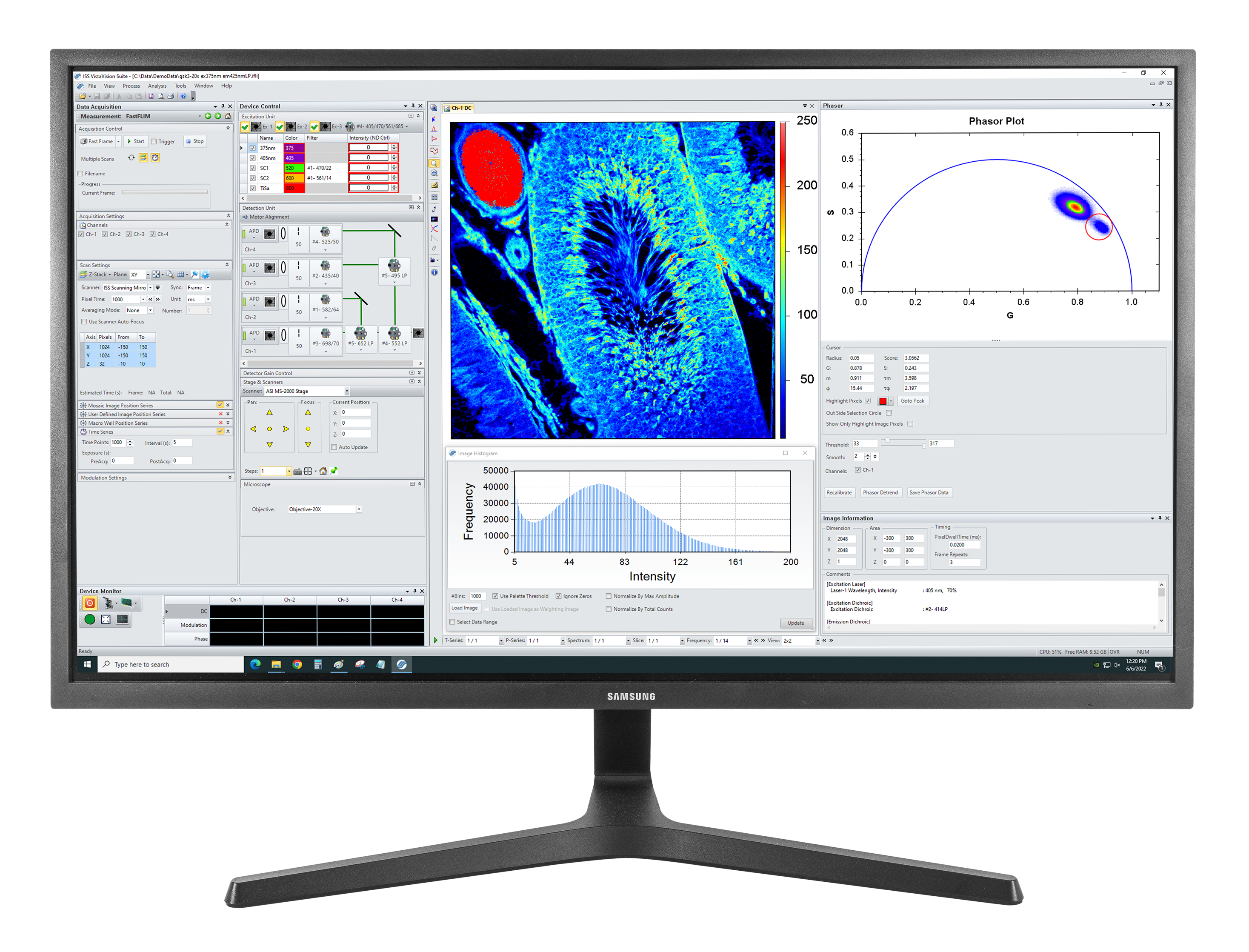
Product Software for Alba STED
VistaVision
VistaVision is a complete software package for confocal microscopy applications including instrument control, data acquisition and data processing. Easy to use, the software has been developed in modular components that can be activated when a specific instrument configuration is selected. The modules include:
- FLIM/PLIM Imaging
- FFS
- smFRET
- Particle Tracking
Product Resources
- Correlative confocal fluorescence lifetime and Atomic Force Microscopy imaging by ISS and JPK
- FLIM Analysis using the Phasor Plots
- Particle Tracking in a 2-Photon Excitation Microscope
- The Sweet PIE (Pulsed Interleaved Excitation)
- Using Alba with the FemtoFiber laser by Toptica for 2-photon quantitative imaging
- FastFLIM STED for Alba v5
- FLIM Analysis Enhanced with Phasor Plotting Aids Quantitative Biology
-
“Spatial resolution enhancement in photon-starved STED imaging using deep learning-based fluorescence lifetime analysis.” Chen, Y.-I., Chang, Y.-J., Sun, Y., Liao, S.-C., Santacruz, S.R. & Yeh, H.-C. Nanoscale, 15(21), pp. 9449–9456, 2023. doi: 10.1039/d3nr00305a.
-
“Epigenetic alterations associated with dexamethasone sodium phosphate through DNMT and TET in RPE cells.” Liu, W., Mohan, S. P., Nagaraj, N.R., Jaganathan, S.S., Wen, Y., Ramasubramanyan, S., & Irudayaraj, J. Molecular Vision, 2021, 27:643-655.
-
“Perfluorooctanoic acid (PFOA) exposure inhibits DNA methyltransferase activities and alters constitutive heterochromatin organization.” Liu, W. & Irudayaraj, J. Food and Chemical Toxicology, 141, p. 111358, 2020, Jul. doi: 10.1016/j.fct.2020.111358.
-
“Monomeric cohesin state revealed by live-cell single-molecule spectroscopy.” Liu, W., Biton, E., Pathania, A., Matityahu, A., Irudayaraj, J. & Onn, I. EMBO reports, 21(2), 2019, Dec. doi: 10.15252/embr.201948211.
-
“Photon-separation to enhance the spatial resolution of pulsed STED microscopy.” Tortarolo, G., Sun, Y., Teng, K.W., Ishitsuka, Y., Lanzanó, L., Selvin, P.R., Barbieri, B., Diaspro, A. & Vicidomini, G. Nanoscale, 11(4), pp. 1754–1761, 2019, Dec. doi: 10.1039/c8nr07485b.
-
“A straightforward STED-background corrected fitting model for unbiased STED-FCS analyses.” Wang, R., Brustlein, S., Mailfert, S., Fabre, R., Fallet, M., Sivankutty, S., Rigneault, H. & Marguet, D. Methods, 140-141(4), pp. 212–222, 2018, May. doi: 10.1016/j.ymeth.2018.02.010.
-
“A novel pulsed STED microscopy method using FastFLIM and the phasor plots.” Sun, Y., Tortarolo, G., Teng, K.-W., Ishitsuka, Y., Coskun, U.C., Liao, S.-C.J., Diaspro, A., Vicidomini, G., Selvin, P.R. & Barbieri, B. Methods, 140-141(4), pp. 212–222, 2017, Feb. doi: 10.1117/12.2267880.