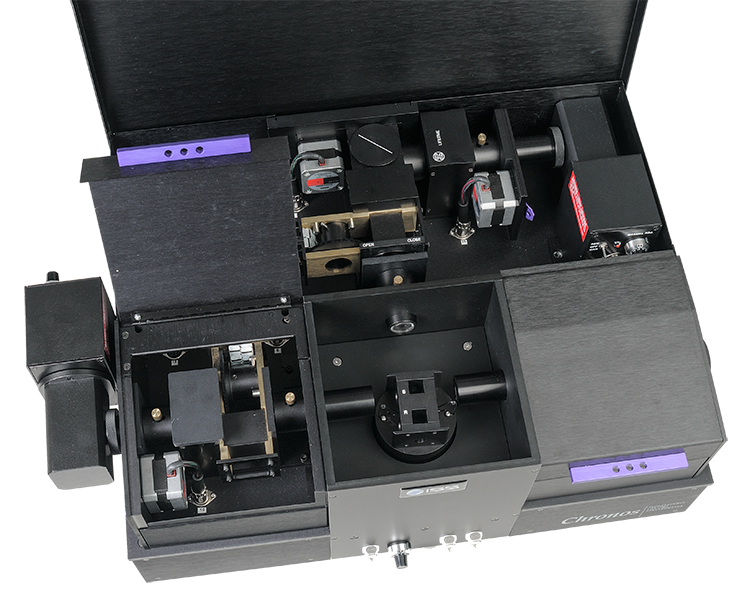
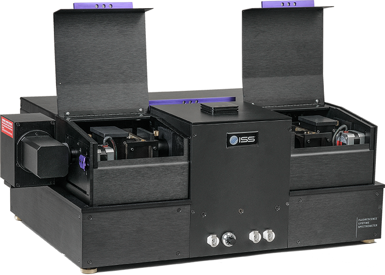


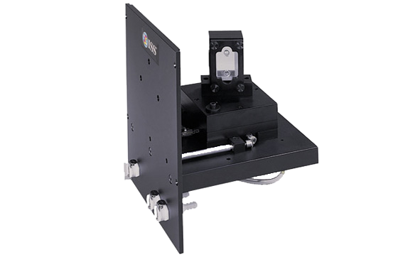
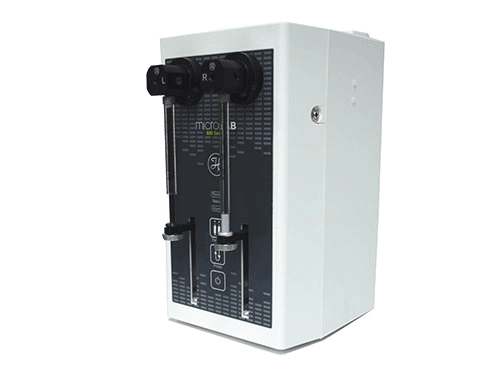
One-Cuvette Sample Compartment

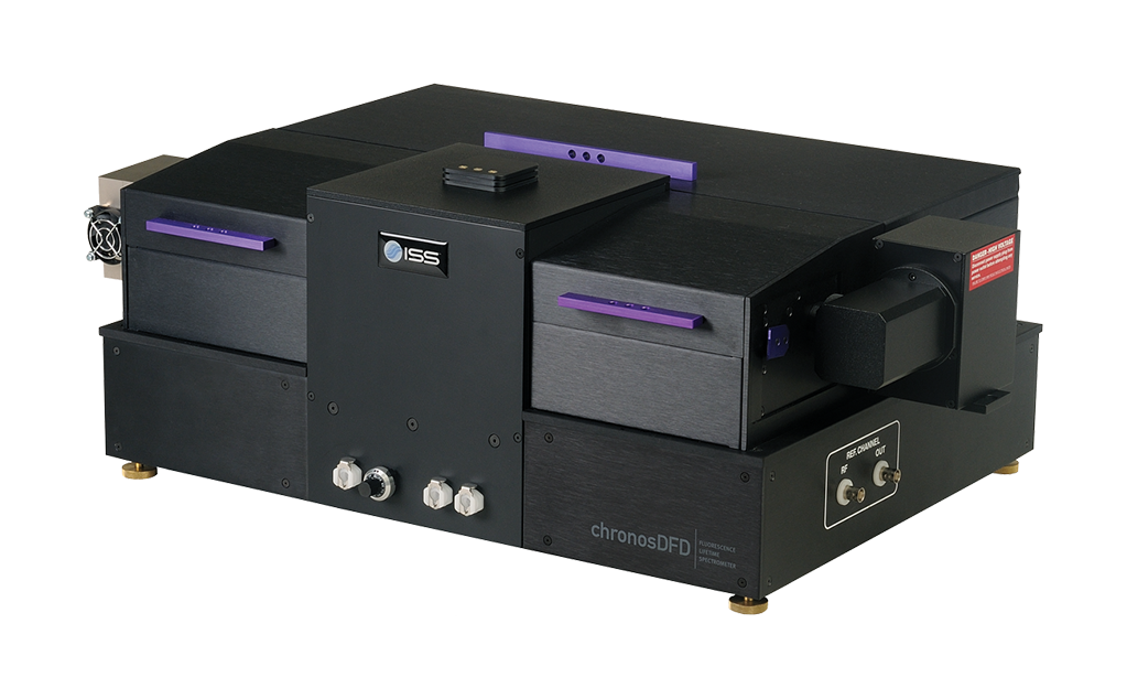
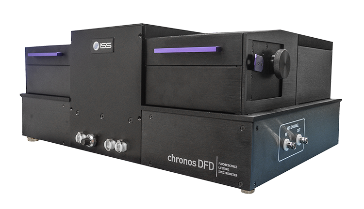
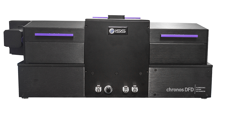
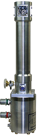
The ChronosDFD is a high-performance digital frequency-domain (DFD) spectrometer designed for rapid and precise fluorescence lifetime measurements, even in complex decay systems. Engineered for efficiency, the system delivers accurate lifetime data in under one second for routine samples, making it ideal for both high-throughput environments and advanced research applications.
At the core of the ChronosDFD is a T-format optical geometry that enables simultaneous acquisition across two emission channels, enhancing data quality and experimental flexibility. The system supports a wide range of excitation sources, including modulated and pulsed laser diodes, supercontinuum lasers, and multiphoton lasers, allowing users to tailor configurations to diverse experimental needs.
Fully automated operation streamlines workflow and ensures reproducibility. Users can easily define custom acquisition protocols and run experiments with minimal intervention, freeing time for analysis and interpretation.
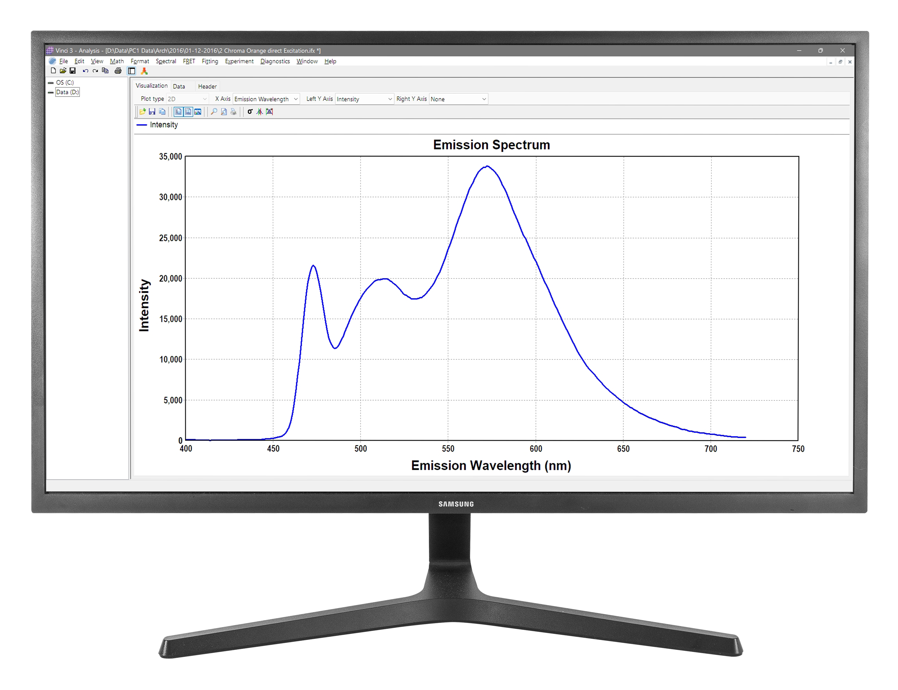
The instrument is powered by Vinci Multidimensional Fluorescence Spectroscopy software, a robust and intuitive Windows-based platform that integrates acquisition, visualization, and analysis within a single environment. Vinci supports a comprehensive suite of measurements, including excitation and emission spectra, polarization and anisotropy, synchronous luminescence, fast and slow kinetics, fluorescence lifetimes, and rotational correlation times. It also enables multidimensional datasets spanning wavelength, time, temperature, polarization, and lifetime domains.
Data are stored in accessible ASCII format with full experimental metadata, ensuring transparency and compatibility with external workflows. Vinci’s analysis capabilities include spectral manipulation, smoothing, corrections, derivatives, and integration, along with advanced fitting routines for multi-exponential and non-exponential decay models, lifetime distributions, and rotational dynamics. Users can also implement custom analysis models with χ² minimization for specialized applications.
Advanced visualization tools provide interactive 2D and 3D plotting, color mapping of user-defined functions, and full control over zooming and rotation. Publication-ready graphics can be exported in standard formats for seamless reporting and presentation.

Frequency Domain Measurements

Maximum Sensitivity

Fully Automated

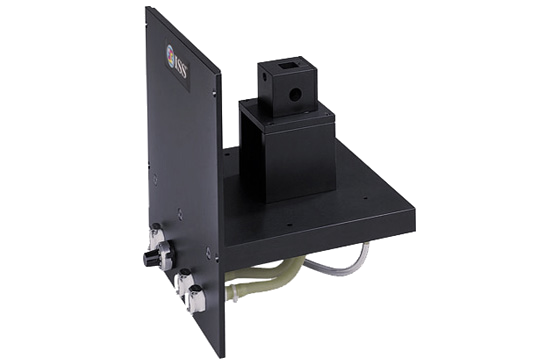
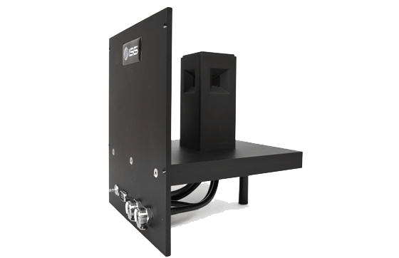
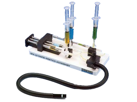
Integration of External Devices

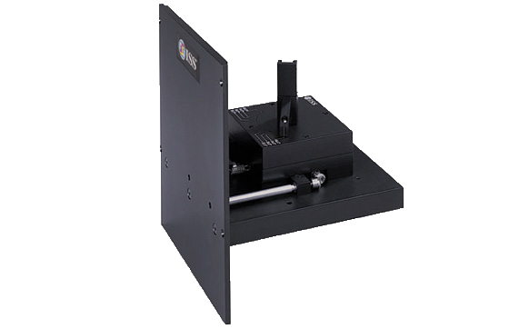
Upgradeable


A comprehensive multidimensional fluorescence spectroscopy software program designed to enhance the capabilities and performance of ISS spectrofluorometers.
Learn More