Overview of PL1
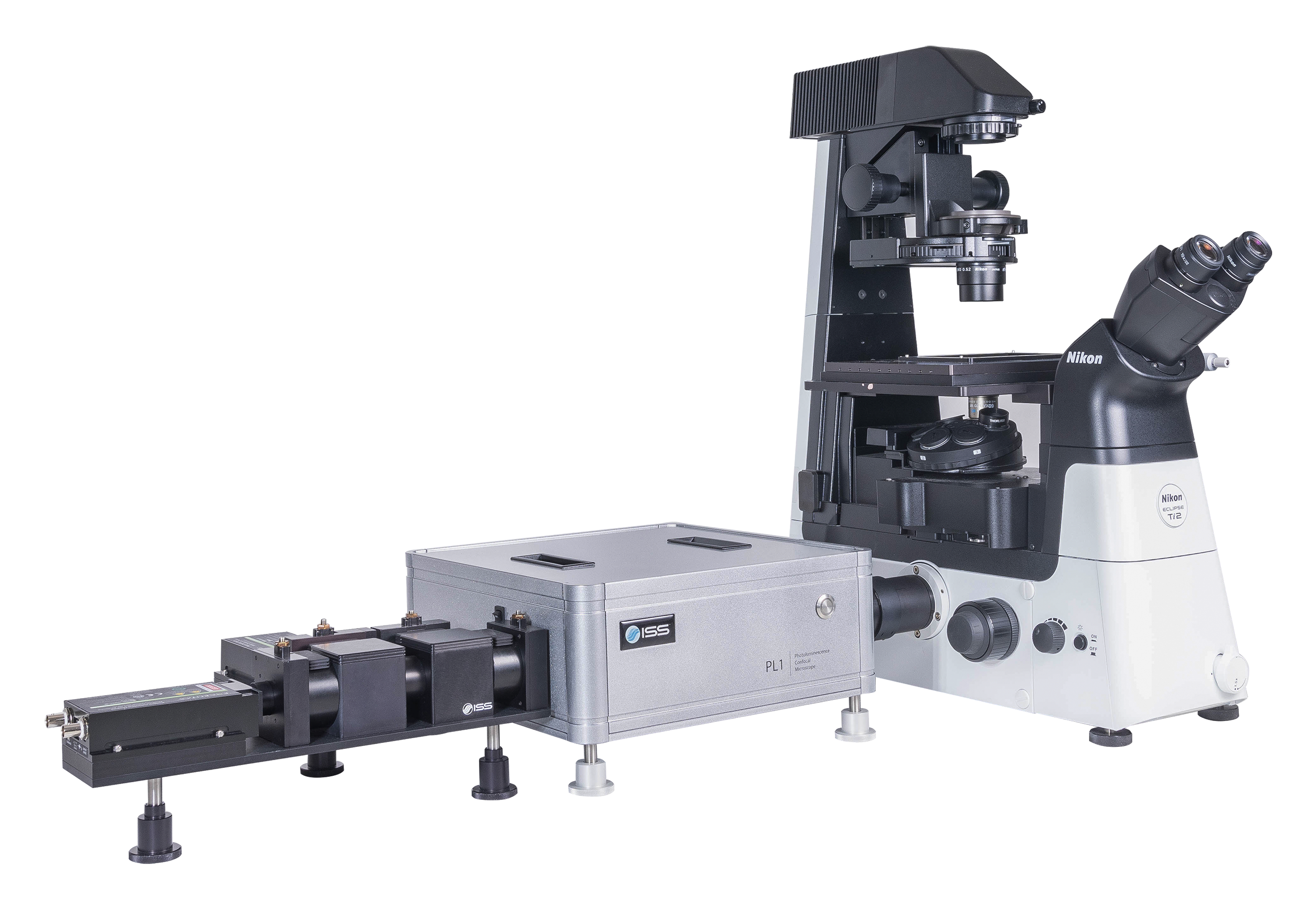
A time-resolved confocal microscope, the PL1 is designed primarily for material sciences research requiring the ultimate sensitivity in FLIM acquisition of large area samples. The sample, with dimensions up to 100 x 100 mm for the upright microscope and up to 120 x 75 mm for the inverted type, is placed on the high-precision, computer-controlled, XY stage that travels from pixel to pixel featuring a 22 nm resolution.
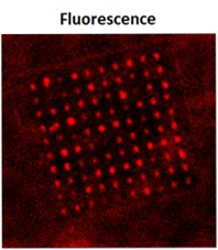
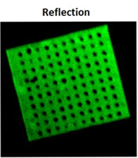
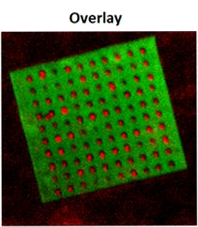
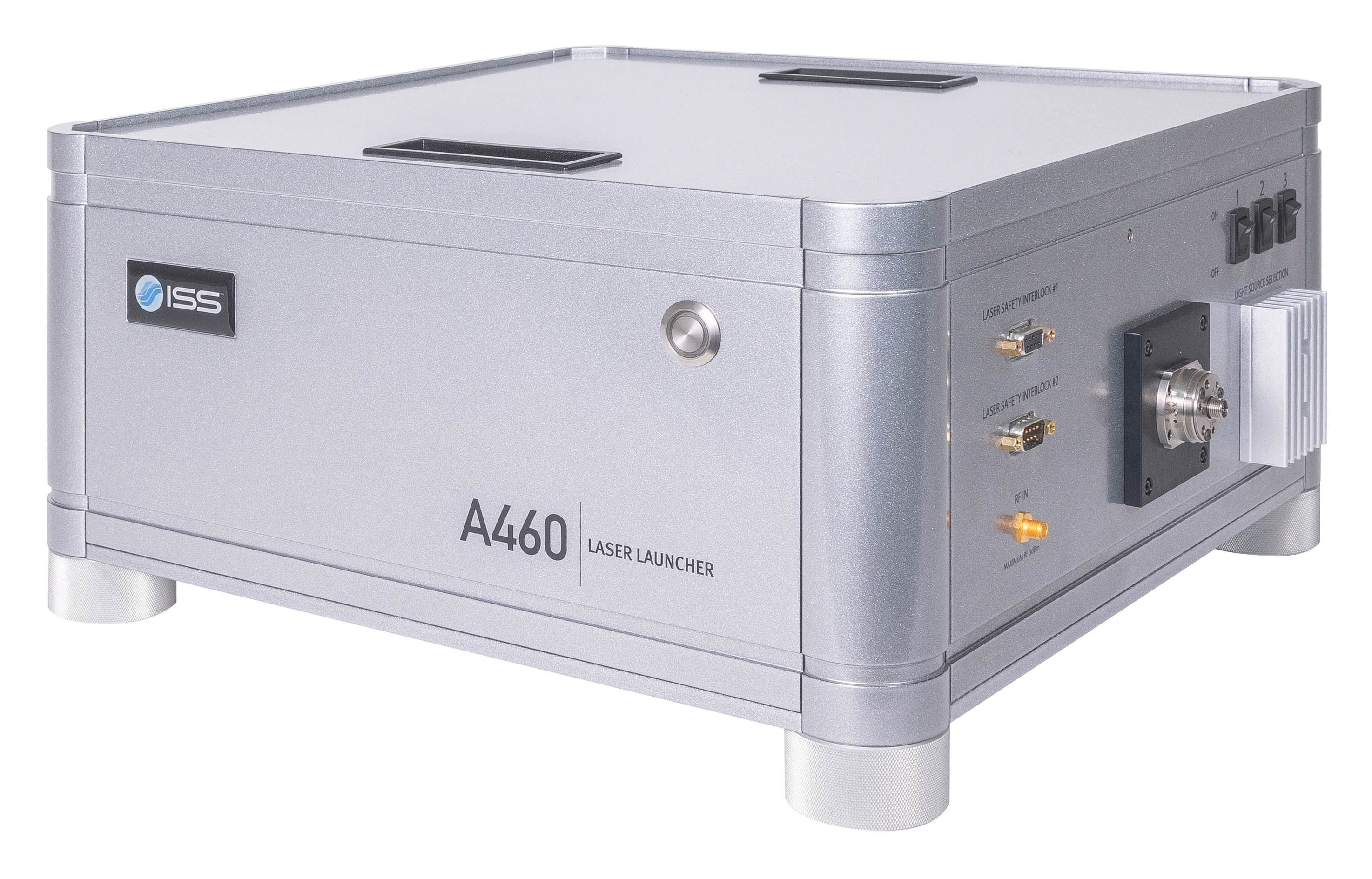
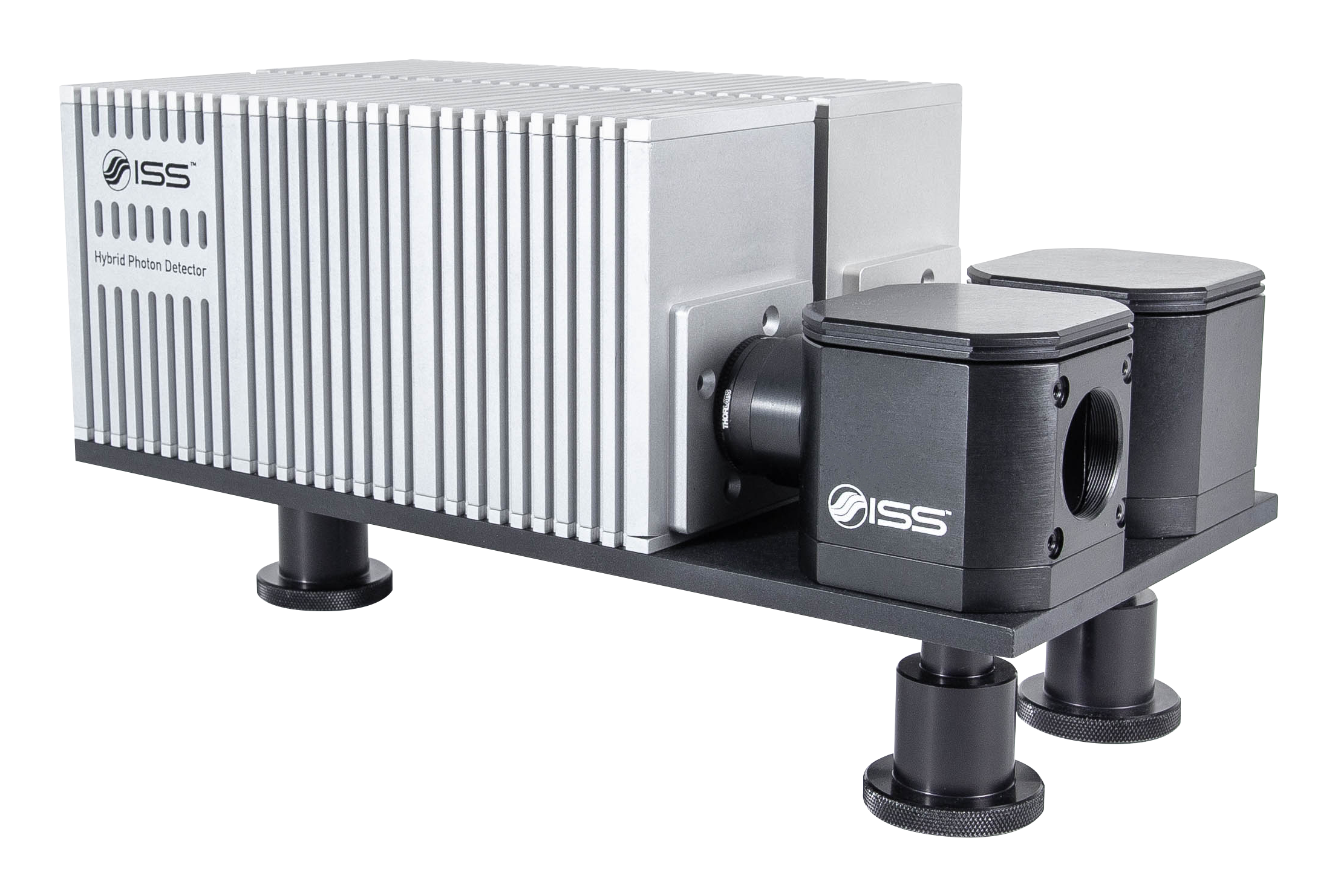
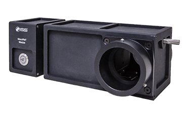
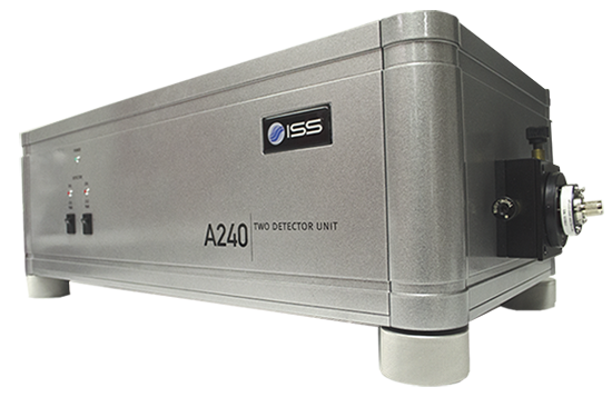
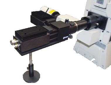
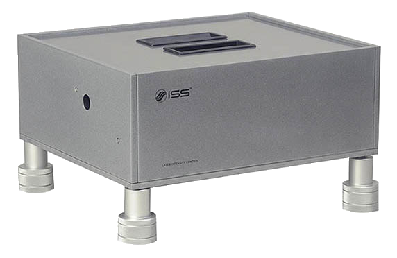
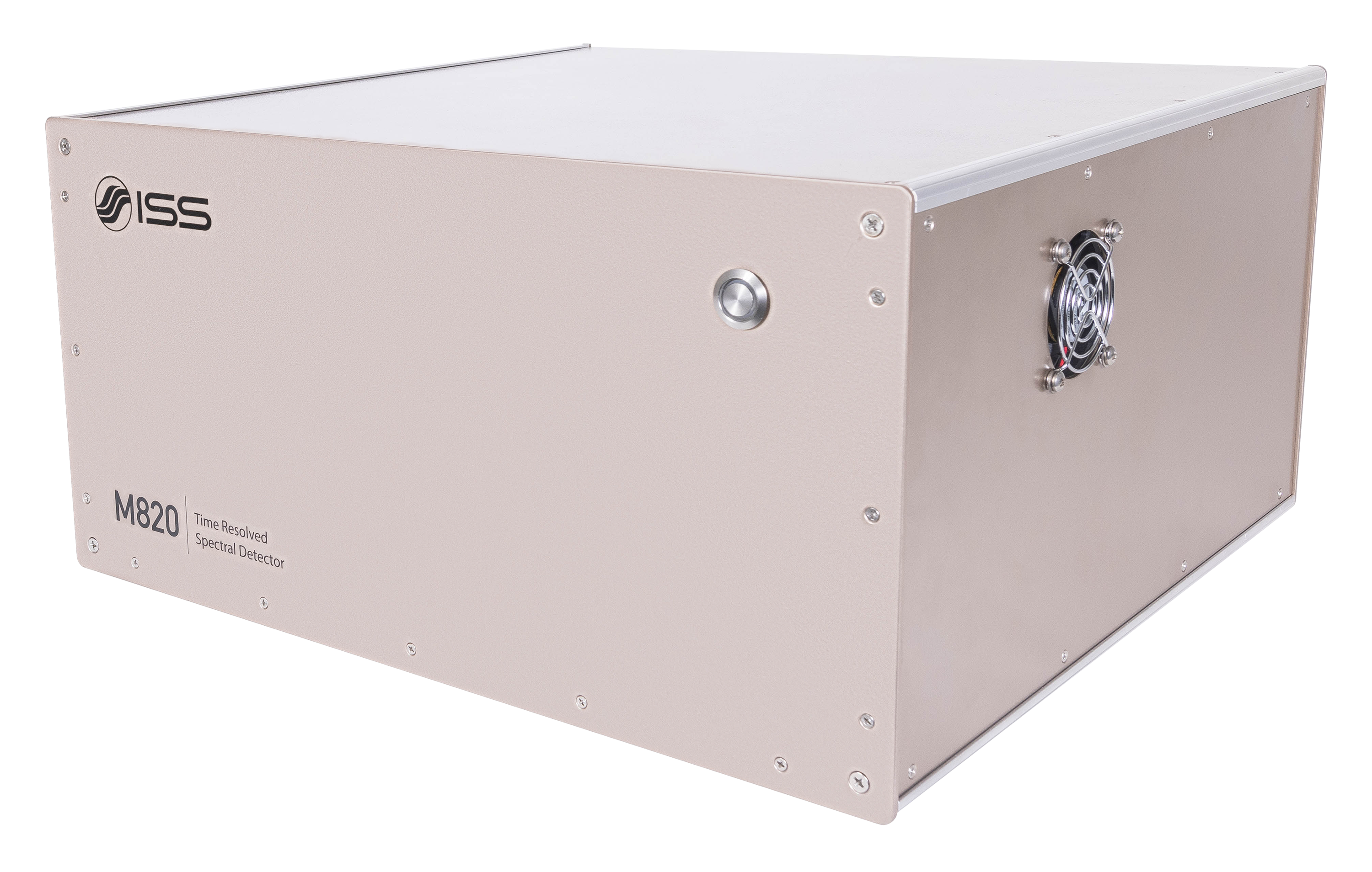
The excitation source is a laser diode, a pulsed laser, or a multiphoton laser. The fluorescence is collected by one detector covering the range 350 – 1050 nm; additional detectors can be added including a spectrograph for pixel spectral acquisition.
Key Features of PL1
FLIM of Large Areas
Upright Microscope:
100 mm x 100 mm
Inverted Microscope:
120 mm x 75 mm
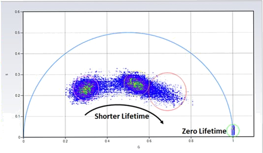
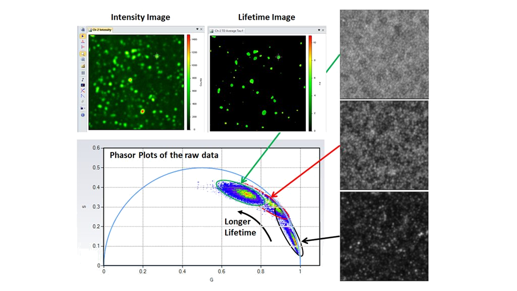
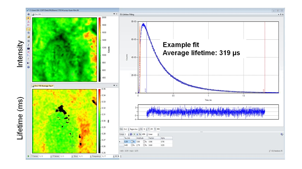
Lifetime Measurements
From 100 ps to 100 ms
Modularity
A selection of laser wavelengths, detectors, number of detection channels, & microscopes.
The Data is Clear!


(Courtesy of Wenjie Liu and Dr. Yaowu Hu; Purdue University; West Lafayette, IN; USA)

(Courtesy of Dr. Xiaodan Zhang; Nan Kai University, Tianjin, P.R. China)

(Courtesy of Dr. Yan Guan, Peking University, Beijing; P.R. China)
Product Specifications for PL1
Microscope and Coupling
- Frame format: Upright or Inverted research microscope
- Magnification: 10X and 60X, oil immersion objective (standard); optional: from 2X to 100X
- Spatial Resolution: diffraction limited
- Eye Observation: bright field by 10X eyepiece with diopter adjustment, field of view: 22 mm
- Imaging Modes:
- Transmission mode: HAL Köhler illumination for bright field imaging by a CMOS camera with options for phase contract and DIC
- Confocal Photoluminescence imaging: laser illumination, single point or scan
XYZ Stage Scan
Closed-loop DC servo control
- XY axis range of travel: 100 mm x 100 mm (upright), 120 mm x 75 mm (inverted)
- XY axis: Resolution = 22 nm, max velocity = 7 mm/s, RMS repeatability
- Z axis: Resolution = 50 nm, maximum velocity = 0.6 mm/s, repeatability = 100 nm
Laser Sources
- CW or pulsed diode laser, repetition rate up to 80 MHz (tunable by software)
- The laser launcher can accommodate up to 3 lasers, ranging from 375 nm to 980 nm
- Each laser has its own intensity control and shutter (operated by software)
Data Acquisition Unit FastFLIM
- Lifetime measurement range: from 100 ps to 100 ms
- Data Acquisition Mode: Photon mode, Time-tagged mode, Time-resolved time-tagged mode (TTTR)
- Dead Time: 3.125 ns, Up to 60 x 106 counts/second
- Computer Connection: USB
Detectors
- Dark Counts < 100/sec; TTS < 350 ps;
- Wavelength Range: 350 - 1050 nm;
- Quantum Efficiency > 70% at 700 nm
Scan Modes
- X, XY, XZ, XYZ, t, Xt, XYt, XZt, XYZt
- Image Format Other than the proprietary file formats which contain the imaging parameters information, VistaVision also supports exporting the acquired data in various formats including JPEG, TIFF, PNG, and AVI.
- Image Processing and Analysis Visualization by various look–up tables, contrasting, thresholding, smoothing, filtering, scaling, statistical analysis by histogram or online profiling.
Lifetime Data Analysis Routines
- Non-linear least square constrained deconvolution fitting routines based on the Marquardt-Levenberg minimization algorithm in both time and frequency domains
- Model-free phasor plots approach for instant and unbiased results
Software
- VistaVision
Computer & Monitor
- High-performance Processor, 32 GB RAM, Windows 10, 64-bit
- 32" monitor, 2556 x 1440 resolution
Power Requirements
- Universal power input: 110 - 240 V, 50/60 Hz, 100 VAC
Measurements for PL1
Intensity and Lifetime Imaging
Fluorescence Fluctuations Spectroscopy (FFS)
Single Molecule FRET Bursts Analysis
Configuration for PL1
Product Software for PL1

VistaVision
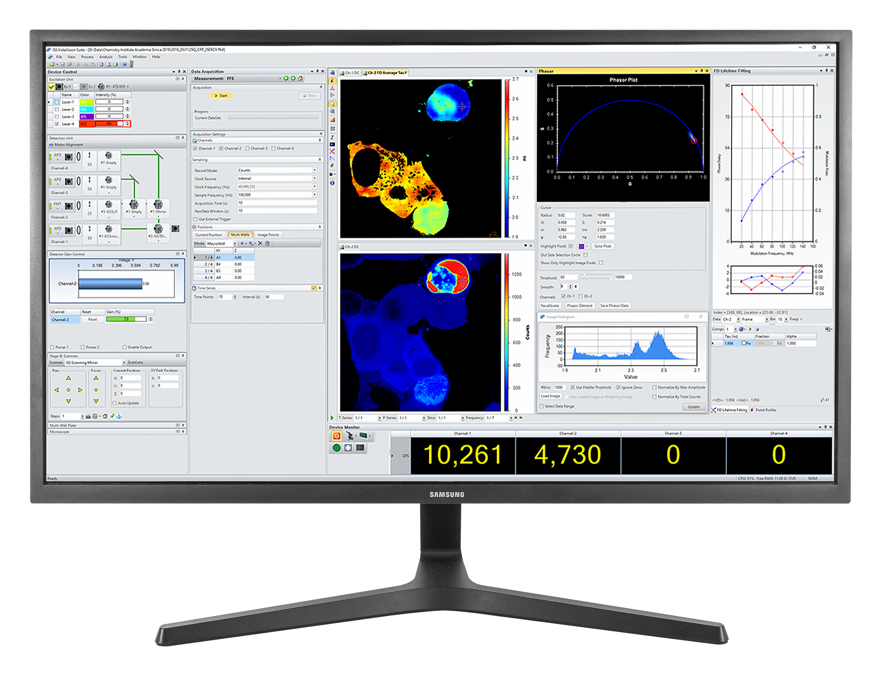
VistaVision is a complete software package for confocal microscopy applications including instrument control, data acquisition and data processing. Easy to use, the software has been developed in modular components that can be activated when a specific instrument configuration is selected. The modules include:
- FLIM/PLIM Imaging
- FFS
- smFRET
- Particle Tracking
Product Resources
-
Correlative confocal fluorescence lifetime and Atomic Force Microscopy imaging by ISS and JPK
-
FLIM Analysis using the Phasor Plots
-
Particle Tracking in a 2-Photon Excitation Microscope
-
The Sweet PIE (Pulsed Interleaved Excitation)
-
Using Alba with the FemtoFiber laser by Toptica for 2-photon quantitative imaging
-
FastFLIM STED for Alba v5