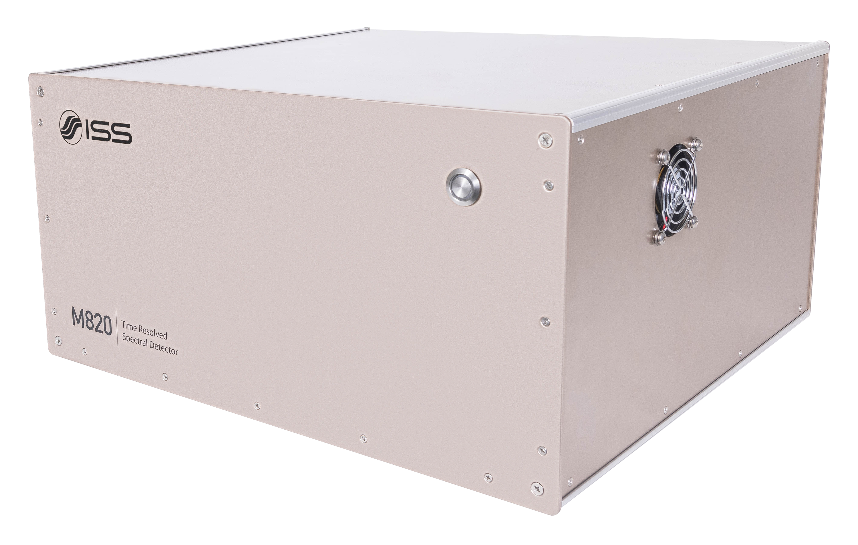
Overview of PL1
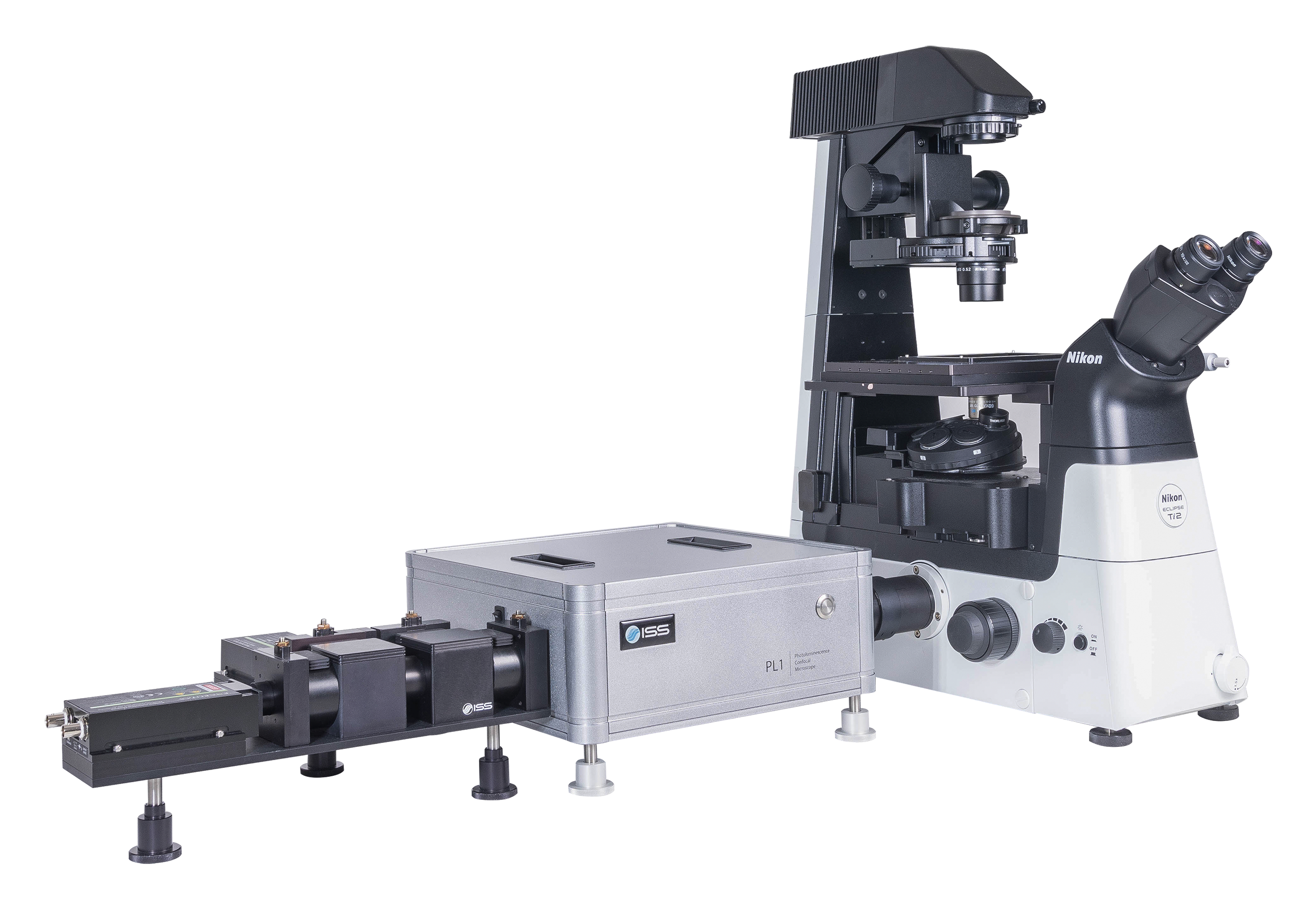
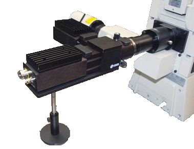
A time-resolved confocal microscope, the PL1 is designed primarily for material sciences research requiring the ultimate sensitivity in FLIM acquisition of large area samples. The sample, with dimensions up to 100 x 100 mm for the upright microscope and up to 120 x 75 mm for the inverted type, is placed on the high-precision, computer-controlled, XY stage that travels from pixel to pixel featuring a 22 nm resolution.
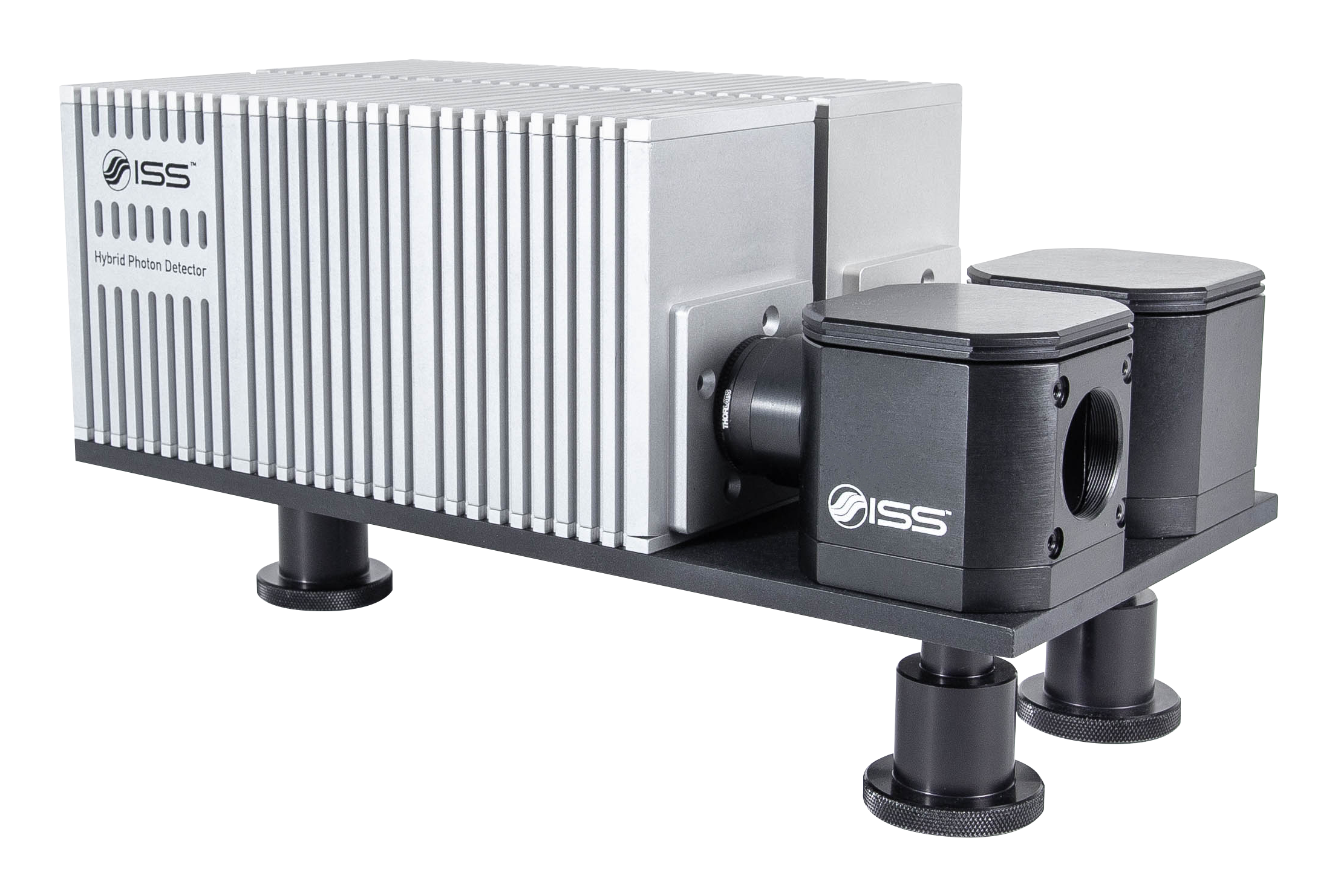
The excitation source is a laser diode, a pulsed laser, or a multiphoton laser. The fluorescence is collected by one detector covering the range 350 – 1050 nm; additional detectors can be added including a spectrograph for pixel spectral acquisition.
Key Features of PL1

FLIM of Large Areas
Upright Microscope:
100 mm x 100 mm
Inverted Microscope:
120 mm x 75 mm

Lifetime Measurements
From 100 ps to 100 ms

Modularity
A selection of laser wavelengths, detectors, number of detection channels, & microscopes.
The Data is Clear!
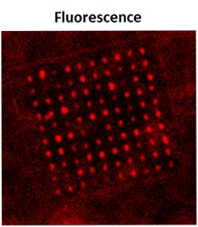
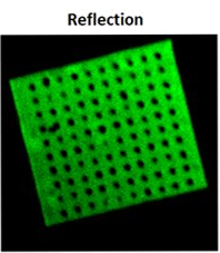
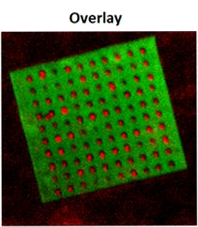
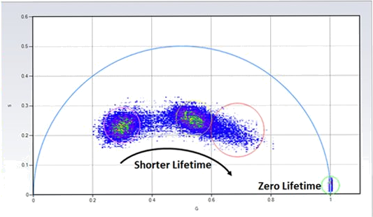
(Courtesy of Wenjie Liu and Dr. Yaowu Hu; Purdue University; West Lafayette, IN; USA)
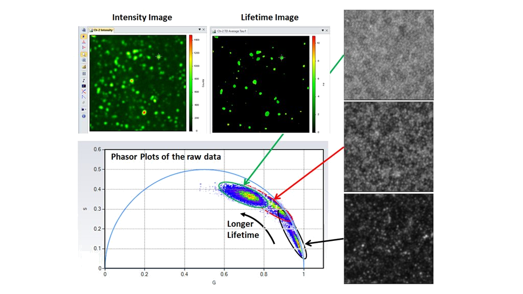
(Courtesy of Dr. Xiaodan Zhang; Nan Kai University, Tianjin, P.R. China)
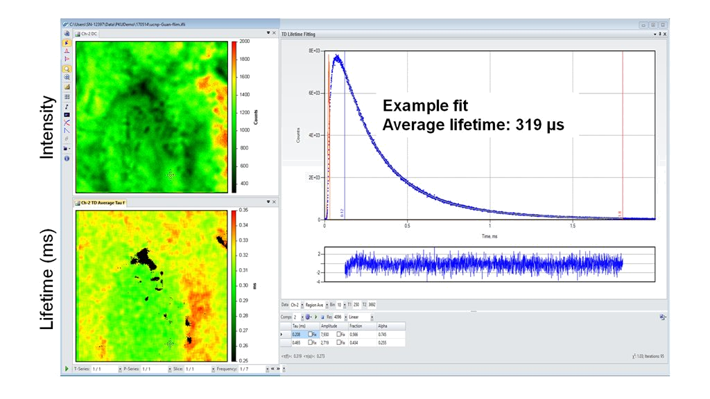
(Courtesy of Dr. Yan Guan, Peking University, Beijing; P.R. China)
Product Specifications for PL1
Microscope and Coupling
- Frame format: Upright or Inverted research microscope
- Magnification: 10X and 60X, oil immersion objective (standard); optional: from 2X to 100X
- Spatial Resolution: diffraction limited
- Eye Observation: bright field by 10X eyepiece with diopter adjustment, field of view: 22 mm
- Imaging Modes:
- Transmission mode: HAL Köhler illumination for bright field imaging by a CMOS camera with options for phase contract and DIC
- Confocal Photoluminescence imaging: laser illumination, single point or scan
XYZ Stage Scan
Closed-loop DC servo control
- XY axis range of travel: 100 mm x 100 mm (upright), 120 mm x 75 mm (inverted)
- XY axis: Resolution = 22 nm, max velocity = 7 mm/s, RMS repeatability
- Z axis: Resolution = 50 nm, maximum velocity = 0.6 mm/s, repeatability = 100 nm
Laser Sources
- CW or pulsed diode laser, repetition rate up to 80 MHz (tunable by software)
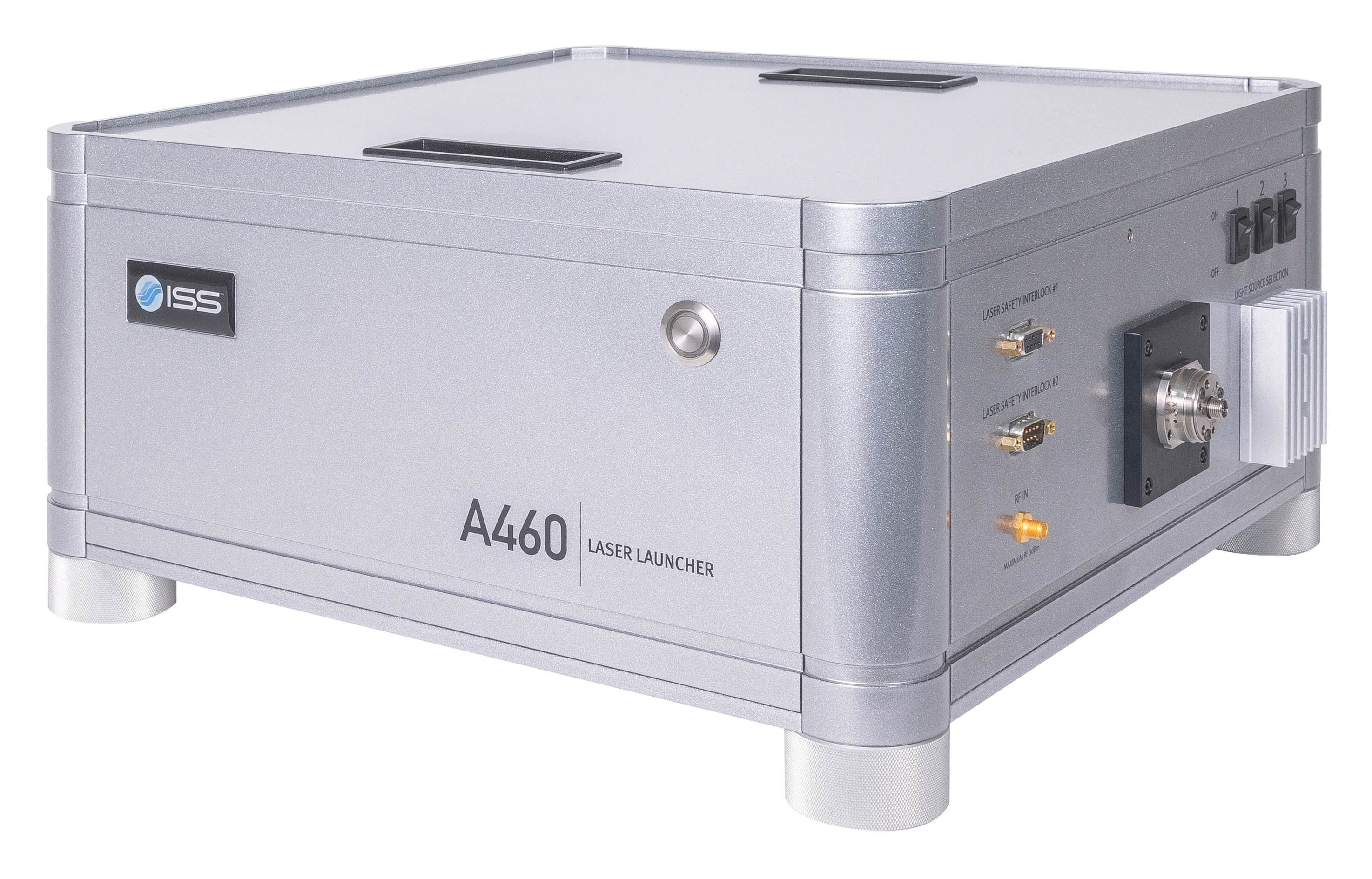
- The laser launcher can accommodate up to 3 lasers, ranging from 375 nm to 980 nm
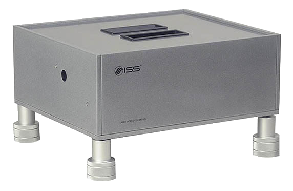
- Each laser has its own intensity control and shutter (operated by software)
Data Acquisition Unit FastFLIM
- Lifetime measurement range: from 100 ps to 100 ms
- Data Acquisition Mode: Photon mode, Time-tagged mode, Time-resolved time-tagged mode (TTTR)
- Dead Time: 3.125 ns, Up to 60 x 106 counts/second
- Computer Connection: USB
Detectors
- Dark Counts < 100/sec; TTS < 350 ps;
- Wavelength Range: 350 - 1050 nm;
- Quantum Efficiency > 70% at 700 nm
Scan Modes
- X, XY, XZ, XYZ, t, Xt, XYt, XZt, XYZt
- Image Format Other than the proprietary file formats which contain the imaging parameters information, VistaVision also supports exporting the acquired data in various formats including JPEG, TIFF, PNG, and AVI.
- Image Processing and Analysis Visualization by various look–up tables, contrasting, thresholding, smoothing, filtering, scaling, statistical analysis by histogram or online profiling.
Lifetime Data Analysis Routines
- Non-linear least square constrained deconvolution fitting routines based on the Marquardt-Levenberg minimization algorithm in both time and frequency domains
- Model-free phasor plots approach for instant and unbiased results
Software
- VistaVision
Computer & Monitor
- High-performance Processor, 32 GB RAM, Windows 11, 64-bit
- 32" monitor, 2556 x 1440 resolution
Power Requirements
- Universal power input: 110 - 240 V, 50/60 Hz, 100 VAC
Measurements for PL1
Intensity and Lifetime Imaging
Fluorescence Fluctuations Spectroscopy (FFS)
Single Molecule FRET Bursts Analysis
Example Configuration for PL1

Product Accessories for PL1
Product Software for PL1
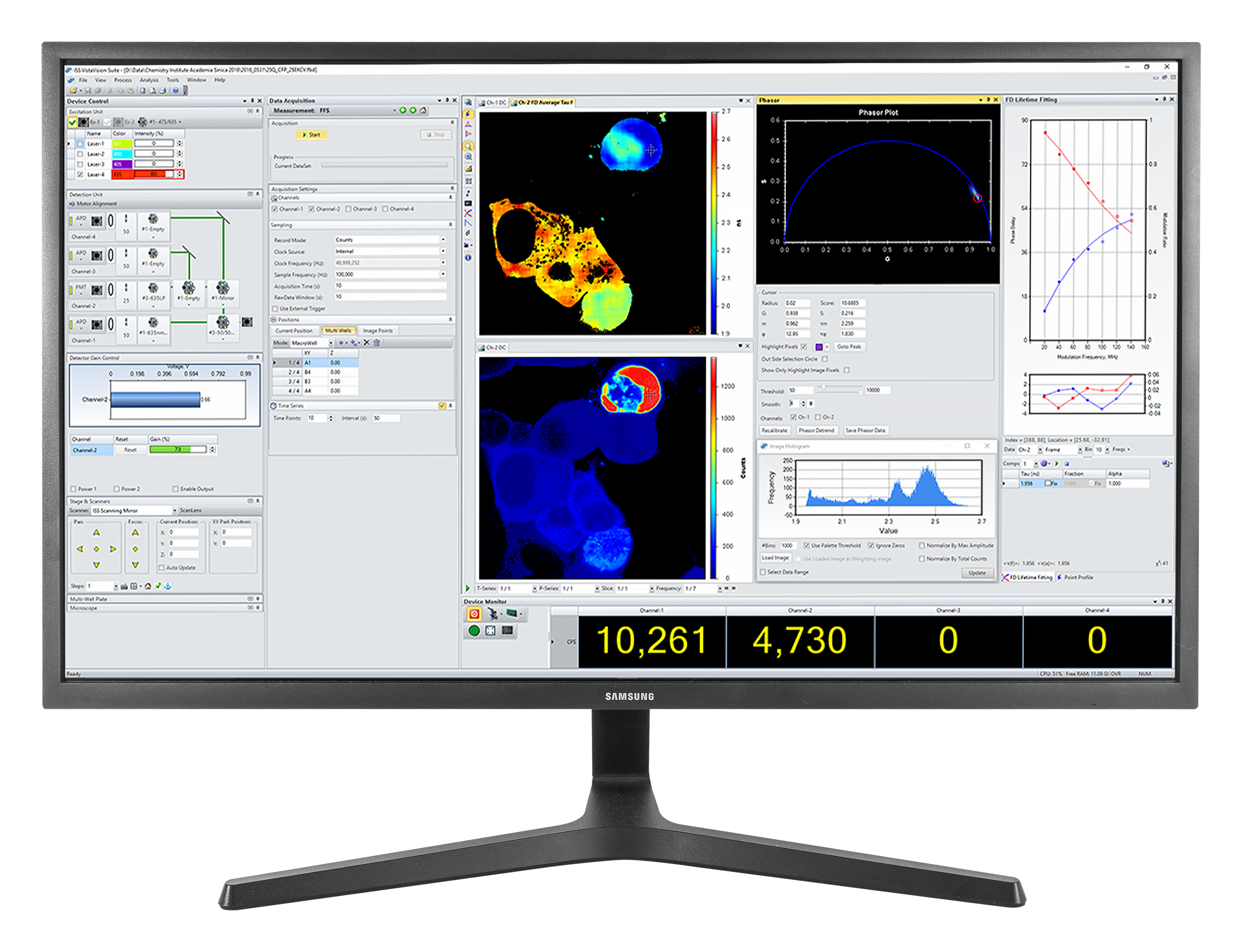
VistaVision
VistaVision is a complete software package for confocal microscopy applications including instrument control, data acquisition and data processing. Easy to use, the software has been developed in modular components that can be activated when a specific instrument configuration is selected. The modules include:
- FLIM/PLIM Imaging
- FFS
- smFRET
- Particle Tracking
Product Resources
-
Correlative confocal fluorescence lifetime and Atomic Force Microscopy imaging by ISS and JPK
-
FLIM Analysis using the Phasor Plots
-
Particle Tracking in a 2-Photon Excitation Microscope
-
The Sweet PIE (Pulsed Interleaved Excitation)
-
Using Alba with the FemtoFiber laser by Toptica for 2-photon quantitative imaging
-
FastFLIM STED for Alba v5
-
“Correlating Photophysical Properties with Stereochemical Expression of 6s2 Lone Pairs in Two-dimensional Lead Halide Perovskites.” Gu, J., Tao, Y., Fu, T., Guo, S., Jiang, X., Guan, Y., Li, X., Li, C., Lü, X. & Fu, Y. Angewandte Chemie International Edition, 62(30), 2023, Jun. doi: 10.1002/anie.202304515.
-
“Phonon-assisted upconversion in twisted two-dimensional semiconductors.” Dai, Y., Qi, P., Tao, G., Yao, G., Shi, B., Liu, Z., Liu, Z., He, X., Peng, P., Dang, Z., Zheng, L., Zhang, T., Gong, Y., Guan, Y., Liu, K. & Fang, Z. Light: Science & Applications, 12(1), 2023, Jan. doi: 10.1038/s41377-022-01051-9.
-
“Color-tunable persistent luminescence in 1D zinc–organic halide microcrystals for single-component white light and temperature-gating optical waveguides.” Zhou, B. & Yan, D. Chemical Science, 13(25), pp. 7429–7436, 2022. doi: 10.1039/d2sc01947g.
-
“Stereochemically Active Lone Pairs and Nonlinear Optical Properties of Two-Dimensional Multilayered Tin and Germanium Iodide Perovskites.” Li, X., Guan, Y., Li, X. & Fu, Y. Journal of the American Chemical Society, 144(39), pp. 18030–18042, 2022, Sep. doi: 10.1021/jacs.2c07535.
-
“Quasi-2D Ruddlesden–Popper Perovskites with Low Trap-States for High Performance Flexible Self-Powered Ultraviolet Photodetectors.” Han, J., Liu, C., Zhang, Y., Guan, Y., Zhang, X., Yu, W., Tang, Z., Liang, Y., Wu, C., Zheng, S. & Xiao, L. Advanced Optical Materials, 10(23), 2022, Sep. doi: 10.1002/adom.202201431.
-
“Achieving Small Temperature Coefficients in Carbon-Based Perovskite Solar Cells by Enhancing Electron Extraction.” Zhang, X., Guan, Y., Zhang, Y., Yu, W., Wu, C., Han, J., Zhang, Y., Chen, C., Zheng, S. & Xiao, L. Advanced Optical Materials, 10(23), p. 2201598, 2022, Sep. doi: 10.1002/adom.202201598.
-
“Red-Emissive Organic Room-Temperature Phosphorescence Material for Time-Resolved Luminescence Bioimaging.” Dai, W., Zhang, Y., Wu, X., Guo, S., Ma, J., Shi, J., Tong, B., Cai, Z., Xie, H. & Dong, Y. CCS Chemistry, 4(8), pp. 2550–2559, 2022, Aug. doi: 10.31635/ccschem.021.202101120.
-
“Understanding Electron–Phonon Interactions in 3D Lead Halide Perovskites from the Stereochemical Expression of 6s2 Lone Pairs.” Huang, X., Li, X., Tao, Y., Guo, S., Gu, J., Hong, H., Yao, Y., Guan, Y., Gao, Y., Li, C., Lü, X. & Fu, Y. Journal of the American Chemical Society, 144(27), pp. 12247–12260, 2022, Jun. doi: 10.1021/jacs.2c03443.
-
“Functionalized SnO2 films by using EDTA-2~M for high efficiency perovskite solar cells with efficiency over 23%.” Tao, J., Liu, X., Shen, J., Wang, H., Xue, J., Su, C., Guo, H., Fu, G., Kong, W. & Yang, S. Chemical Engineering Journal, 430, p. 132683, 2022, Feb. doi: 10.1016/j.cej.2021.132683.
-
“Chemical Polishing of Perovskite Surface Enhances Photovoltaic Performances.” Zhao, L., Li, Q., Hou, C.-H., Li, S., Yang, X., Wu, J., Zhang, S., Hu, Q., Wang, Y., Zhang, Y., Jiang, Y., Jia, S., Shyue, J.-J., Russell, T.P., Gong, Q., Hu, X. & Zhu, R. Journal of the American Chemical Society, 144(4), pp. 1700–1708, 2022, Jan. doi: 10.1021/jacs.1c10842.
-
“Depth-dependent defect manipulation in perovskites for high-performance solar cells.” Zhang, Y., Wang, Y., Zhao, L., Yang, X., Hou, C.-H., Wu, J., Su, R., Jia, S., Shyue, J.-J., Luo, D., Chen, P., Yu, M., Li, Q., Li, L., Gong, Q. & Zhu, R. Energy & Environmental Science, 14(12), pp. 6526–6535, 2021. doi: 10.1039/d1ee02287c.
-
“Lead-free Double Perovskite Cs2AgIn0.9Bi0.1Cl6 Quantum Dots for White Light-Emitting Diodes.” Zhang, Y., Zhang, Z., Yu, W., He, Y., Chen, Z., Xiao, L., Shi, J.-j., Guo, X., Wang, S. & Qu, B. Advanced Science, 9(2), p. 2102895, 2021, Nov. doi: 10.1002/advs.202102895.
-
“Additive Engineering for Efficient and Stable MAPbI3-Perovskite Solar Cells with an Efficiency of over 21%.” Tao, J., Wang, Z., Wang, H., Shen, J., Liu, X., Xue, J., Guo, H., Fu, G., Kong, W. & Yang, S. ACS Applied Materials & Interfaces, 13(37), pp. 44451–44459, 2021, Sep. doi: 10.1021/acsami.1c13136.
-
“Lanthanide Upconverted Microlasing: Microlasing Spanning Full Visible Spectrum to Near-Infrared under Low Power, CW Pumping.” Yang, X.-F., Lyu, Z.-Y., Dong, H., Sun, L.-D. & Yan, C.-H. Small, 17(41), 2021, Sep. doi: 10.1002/smll.202103140.
-
“Low-Dimensional Organic Metal Halide Hybrids with Excitation-Dependent Optical Waveguides from Visible to Near-Infrared Emission.” Wu, S., Zhou, B. & Yan, D. ACS Applied Materials & Interfaces, 13(22), pp. 26451–26460, 2021, May. doi: 10.1021/acsami.1c03926.
-
“Near-Unity Cyan-Green Emitting Lead-Free All-Inorganic Cesium Copper Chloride Phosphors for Full-Spectrum White Light-Emitting Diodes.” Bai, W., Shi, S., Lin, T., Zhou, T., Xuan, T. & Xie, R.-J. Advanced Photonics Research, 2(3), p. 2000158, 2021, Feb. doi: 10.1002/adpr.202000158.
-
“Dual-mode excitation β-NaGdF4:Yb/Er@β-NaGdF4:Yb/Nd core–shell nanoparticles with NIR-II emission and 5 nm cores: controlled synthesis via NaF/RE regulation and the growth mechanism.” Cheng, C., Xu, Y., De, G., Wang, J., Wu, W., Tian, Y. & Wang, S. CrystEngComm, 22(38), pp. 6330–6338, 2020. doi: 10.1039/d0ce01113d.
-
“Superior Carrier Lifetimes Exceeding 6 µs in Polycrystalline Halide Perovskites.” Yang, X., Fu, Y., Su, R., Zheng, Y., Zhang, Y., Yang, W., Yu, M., Chen, P., Wang, Y., Wu, J., Luo, D., Tu, Y., Zhao, L., Gong, Q. & Zhu, R. Advanced Materials, 32(39), p. 2002585, 2020, Aug. doi: 10.1002/adma.202002585.
-
“Engineering of Upconverted Metal–Organic Frameworks for Near-Infrared Light-Triggered Combinational Photodynamic/Chemo-/Immunotherapy against Hypoxic Tumors.” Shao, Y., Liu, B., Di, Z., Zhang, G., Sun, L.-D., Li, L. & Yan, C.-H. Journal of the American Chemical Society, 142(8), pp. 3939–3946, 2020, Jan. doi: 10.1021/jacs.9b12788.
-
“Upconversion Lifetime Imaging of Highly-Crystalline Gd-Based Fluoride Nanocrystals Featuring Strong Luminescence Resulting from Multiple Luminescent Centers.” Xu, Y., Zeng, Z., Zhang, D., Liu, S., Wang, X., Li, S., Cheng, C., Wang, J., Liu, Y., De, G., Zhang, C., Qin, W. & Du, Y. Advanced Optical Materials, 8(4), p. 1901495, 2019, Dec. doi: 10.1002/adom.201901495.
-
“Multifunctional Tetracene/Pentacene Host/Guest Nanorods for Enhanced Upconversion Photodynamic Tumor Therapy.” Zhang, R., Guan, Y., Zhu, Z., Lv, H., Li, F., Sun, S. & Li, J. ACS Applied Materials & Interfaces, 11(41), pp. 37479–37490, 2019, Sep. doi: 10.1021/acsami.9b12967.
-
“High Efficiency (16.37%) of Cesium Bromide—Passivated All-Inorganic CsPbI2Br Perovskite Solar Cells.” Zhang, Y., Wu, C., Wang, D., Zhang, Z., Qi, X., Zhu, N., Liu, G., Li, X., Hu, H., Chen, Z., Xiao, L. & Qu, B. Solar RRL, 3(11), p. 1900254, 2019, Aug. doi: 10.1002/solr.201900254.
-
“FAPbI3 Flexible Solar Cells with a Record Efficiency of 19.38% Fabricated in Air via Ligand and Additive Synergetic Process.” Wu, C., Wang, D., Zhang, Y., Gu, F., Liu, G., Zhu, N., Luo, W., Han, D., Guo, X., Qu, B., Wang, S., Bian, Z., Chen, Z. & Xiao, L. Advanced Functional Materials, 29(34), p. 1902974, 2019, Jun. doi: 10.1002/adfm.201902974.
-
“Band-Aligned Polymeric Hole Transport Materials for Extremely Low Energy Loss α-CsPbI3 Perovskite Nanocrystal Solar Cells.” Yuan, J., Ling, X., Yang, D., Li, F., Zhou, S., Shi, J., Qian, Y., Hu, J., Sun, Y., Yang, Y., Gao, X., Duhm, S., Zhang, Q. & Ma, W. Joule, 2(11), pp. 2450–2463, 2018, Nov. doi: 10.1016/j.joule.2018.08.011.
-
“Composition-Graded Cesium Lead Halide Perovskite Nanowires with Tunable Dual-Color Lasing Performance.” Huang, L., Gao, Q., Sun, L.-D., Dong, H., Shi, S., Cai, T., Liao, Q. & Yan, C.-H. Advanced Materials, 30(27), p. 1800596, 2018, May. doi: 10.1002/adma.201800596.
